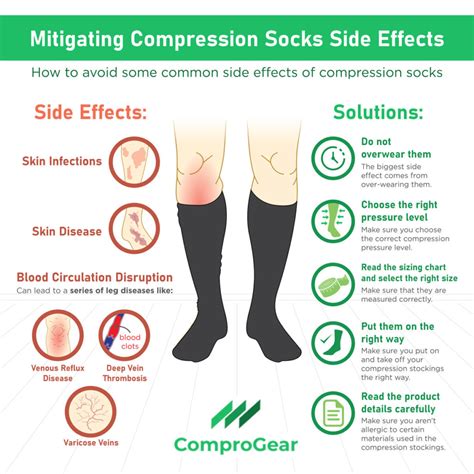
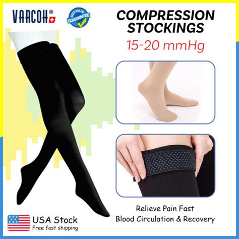
dvt compression test|best compression stockings for dvt : exporter Venous ultrasound is the standard imaging test for patients suspected of having acute deep venous thrombosis (DVT). There is variability and disagreement among authoritative groups regarding the necessary .
Resultado da O Restaurante Guru permite que você descubra ótimos lugares para comer perto da sua localização. Leia os cardápios dos restaurantes e as .
{plog:ftitle_list}
NOTÍCIAS - Joinville Esporte Clube – JOGA JUNTO
Tests used to diagnose or rule out DVT include: D-dimer blood test. D dimer is a type of protein produced by blood clots. Almost all people with severe DVT have increased blood levels of D dimer. This test often can help rule out pulmonary embolism (PE). Duplex ultrasound.Many things can increase the risk of developing deep vein thrombosis (DVT). .
DVT (deep vein thrombosis) prevention and treatment. 44 Replies Tue, Oct 29, 2024 .Warfarin (Jantoven) is a medicine to prevent blood clots. It may save your life .In this section, we will go over a step-by-step ultrasound approach to evaluate for deep vein thrombosis by visualizing the veins and using venous compression using a standard DVT Ultrasound Protocol.
Venous ultrasound is the standard imaging test for patients suspected of having acute deep venous thrombosis (DVT). There is variability and disagreement among authoritative groups regarding the necessary . Two-point compression has been widely accepted as a rapid way to assess for DVT in patients with a low pretest probability, making this an even more rapid way to assess .
D-Dimer testing is sometimes used when DVT is suspected; a negative result helps to exclude DVT, whereas a positive result is nonspecific and requires additional testing to confirm DVT. Treatment is with anticoagulants.
Deep venous thrombosis (DVT) is a major cause of morbidity and is a common presenting complaint to the emergency department (ED). Point-of-care two-point compression .Compression ultrasonography (CUS) is the first-line imaging test in the diagnostic management of suspected deep vein thrombosis (DVT) of the lower extremity. Three CUS strategies are .ABSTRACT: Venous ultrasound is the standard imaging test for patients suspected of having acute deep venous thrombosis (DVT). There is variability and disagreement among .
Compression ultrasonography has proven to be a highly sensitive and specific modality for the recognition of lower extremity DVTs without the need for radiation or contrast exposure. [14,.
DVT in the leg is the most common type of venous thrombosis. However, a clot can form anywhere in the venous system. If a part or all of the blood clot in the vein breaks off . This is the standard test for DVT. Your doctor will run an ultrasound to scan parts of your body for clots in your veins. . Home remedies for DVT. Compression stockings. . Deep vein thrombosis .For the focused deep vein thrombosis (DVT) compression ultrasonographic examination, . Towards evidence based emergency medicine: best BETs from the Manchester Royal Infirmary. Using the ultrasound compression test for . Researchers have extensively studied the lower extremities' deep vein thrombosis (DVT), with the increased use of central venous catheters, cardiac pacemakers/defibrillators, and peripherally inserted central catheter (PICC) lines. . The best way to confirm a diagnosis is with compression duplex ultrasonography. This test has a sensitivity .
Tests Related to DVT. DVT can bring on a dangerous condition called a pulmonary embolism (PE). For some people, a PE is the first sign they have DVT. A pulmonary embolism is when a deep-vein clot .
A deep-vein thrombosis (DVT) is a blood clot that forms within the deep veins, usually of the leg, but can occur in the veins of the arms and the mesenteric and cerebral veins.It is a common disorder and belongs to the venous thromboembolism disorders. DVTs represent the third most common cause of death from cardiovascular disease after heart attacks and stroke, and . The two-point compression group included all patients with DVT locations detected by a two-point compression test. Outcomes The primary outcome of this study was the prevalence and distribution of isolated proximal lower extremity deep venous thrombi in locations other than the common femoral and popliteal veins.Intermittent pneumatic compression (IPC) devices are used to help prevent blood clots in the deep veins of the legs. . Deep vein thrombosis (DVT) is a blood clot that forms in a vein deep inside the body. In most cases, this clot forms inside one of the deep veins of the thigh or lower leg. . Who to call after the test or procedure if you .
Compression ultrasonography (CUS) is the first-line imaging test in the diagnostic management of suspected deep vein thrombosis (DVT) of the lower extremity. Three CUS strategies are used in clinical practice. However, their relative diagnostic .
Deep vein thrombosis in upper extremities: Clinical characteristics, management strategies and long-term outcomes from the COMMAND VTE Registry. . The compression may be due to a normal or an accessory first rib or fibrous band . Contrast venography was the definitive test for the diagnosis of DVT in the past but has been largely replaced .
Remarks: Thrombolysis is reasonable to consider for patients with limb-threatening DVT (phlegmasia cerulea dolens) and for selected younger patients at low risk for bleeding with symptomatic DVT involving the iliac and common femoral veins (higher risk for more severe postthrombotic syndrome [PTS] 3).Patients in these categories who value rapid resolution of .In this 1-minute video POCUS tip, learn how to perform a Venous Compression Test for DVT. Ready to get started on your POCUS journey? Check out our many certificates and certifications here.Bedside ultrasound can be used to conduct compression testing on lower extremity vasculature to assess for DVT; Intended to be rapid, limited, but revealing most clinically significant DVTs; Amongst ED providers, there is a sensitivity of 95% and specificity of 96%; Indications. Clinical suspicion of DVT: edema, tenderness over the calf, Homan .

Venous ultrasound is the standard imaging test for patients suspected of having acute deep venous thrombosis (DVT). There is variability and disagreement among authoritative groups regarding the necessary components of the test. Some protocols include scanning the entire lower extremity, whereas others recommend scans limited to the thigh and knee .Noninvasive diagnosis of deep vein thrombosis (DVT) is based on ultrasound examination of the leg veins, usually restricted to only compression of the proximal veins (CUS). Patients with negative CUS findings require a second examination or a combination with other tests, which impairs clinical effi . Deep vein thrombosis (DVT) is a blood clot that develops within a deep vein in the body. . These include a specific blood test called a D dimer, and/or an ultrasound. An ultrasound scan shows if blood is flowing normally through the veins or if there are any clots. . Compression socks. You’ll be prescribed compression socks to treat DVT .
Compression Stockings for DVT. If you have been diagnosed with deep vein thrombosis (DVT), your doctor may prescribe compression stockings. These stockings help to improve blood flow in the legs and lower . In this 1-minute video POCUS tip, learn how to perform a Venous Compression Test for DVT.Visit Us -- https://www.pocus.org/View Our POCUS Resources -- https:.How is deep vein thrombosis or DVT diagnosed? There are multiple ways healthcare providers can diagnose DVT. . a DVT diagnosis is made. The ability to completely flatten a vein with compression is the most useful way to be certain that a clot is not present. . if the test is negative, the chance that there is a DVT is so small that .
should dvt wear compression socks
If the test shows high levels of the substance, you may have DVT. These tests may be used as a first step to look for signs of a blood clot in otherwise healthy people. Compression ultrasound looks for blood clots in the deep veins of your legs. This test uses sound waves to create pictures of blood flowing in your veins.

Additionally a PubMed search [compression test, DVT, embolism, risk] yielded 0 relevant papers. Comment(s) The lack of evidence in this area is not surprising as a controlled clinical trial would be unlikely to gain ethical approval. However this presents the clinician with the all‐too‐familiar situation of wondering whether or not they . Your healthcare provider will most likely perform a compression ultrasound, but other tests, like a venogram, CT scan, or D-dimer test, can also be used to diagnose DVT. Through a compression ultrasound, your practitioner is able to see the blood clot and the obstruction of blood flow in the vein.ABSTRACT: Venous ultrasound is the standard imaging test for patients suspected of having acute deep venous thrombosis (DVT). There is . of acute DVT. CDUS is compression of the deep veins from the inguinal ligament to the ankle (including pos-terior tibial and peroneal veins in the calf), right and .
Johnson SA, Stevens SM, Woller SC, et al. Risk of deep vein thrombosis following a single negative whole-leg compression ultrasound: a systematic review and meta-analysis. JAMA. 2010;303(5):438-445.
A short cut review was carried out to establish whether patients with deep vein thrombosis (DVT) are at risk of embolism during ultrasound compression testing. No papers were found that directly answered the clinical question. The clinical bottom line is that currently there is no evidence to suggest that compressing vessels in order to identify a DVT could cause an . Keywords: Compression ultrasound, deep-vein thrombosis, emergency department. . Venography is considered the most accurate test, which can confirm or rule out DVT. Even though venography is the gold standard for the diagnosis of DVT, it is invasive and requires contrast agents. On the other hand, duplex ultrasonography (DUS) is a suitable . The point-of-care ultrasound DVT (POCUS DVT) examination can facilitate rapid bedside diagnosis and treatment of lower extremity DVT. Awaiting radiology-performed Doppler ultrasonography and interpretation by radiologists can lead to delays in lifesaving anticoagulation, and the POCUS DVT examination can provide timely diagnostic information in the patient with .Deep vein thrombosis (DVT) is a type of venous thrombosis involving the formation of a blood clot in a deep vein, most commonly in the legs or pelvis. [9] [a] A minority of DVTs occur in the arms. [11]Symptoms can include pain, swelling, redness, and enlarged veins in the affected area, but some DVTs have no symptoms. [1]The most common life-threatening concern with DVT is .
cervical spine tests foraminal compression distraction
cervical spine tests foraminal compression distraction valsalva
雙人遊戲 ‧雙人遊戲 ‧聯機遊戲桃 ‧雙人闖關 ‧雙人格鬥 ‧雙人 .
dvt compression test|best compression stockings for dvt