echo test machine|echo machine hospital : advice An echo test can allow your health care team to look at your heart’s structure and check how well your heart functions. The test helps your health care team find out: 1. The size and shape of your heart, and the size, thickness and movement of your heart’s walls. 2. How . See more Resultado da 37:00. Coroa loira de 53 anos deixa negão abusar da sua buceta rosada 35835 82% 10:00. Coroa brasileira sai com novinho quando marido não .
{plog:ftitle_list}
web29 de jan. de 2024 · A prática de apostas online em solo português é perfeitamente legal, desde que seja feita exclusivamente através de plataformas licenciadas pelo SRIJ, em .
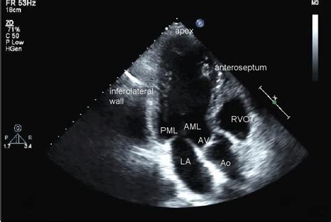
An echocardiogram (echo) uses high frequency sound waves (ultrasound) to make pictures of your heart. The test is also called echocardiography or diagnostic cardiac ultrasound. The types of echocardiograms are: 1. Transthoracic echocardiography 2. Stress echocardiography 3. Transesophageal echocardiography . See more
An echo test can allow your health care team to look at your heart’s structure and check how well your heart functions. The test helps your health care team find out: 1. The size and shape of your heart, and the size, thickness and movement of your heart’s walls. 2. How . See moreEcho tests are done by specially trained technicians. You may have your test done in your doctor’s office, an emergency room, an operating room, a hospital clinic or a hospital room. The test takes about an hour. 1. You lie on a table and small metal disks . See moreSpecially trained technicians conduct echo tests. You may have your test done in a medical office, emergency room, operating room, hospital clinic . See moreYour health care professional will talk with you after looking at your echo pictures and discuss what the pictures show. Download our printable sheet: What is an Echocardiogram? (PDF) See more
An echocardiogram is a test that uses ultrasound to show how your heart muscle and valves are working. These sound waves make moving pictures of your heart so your doctor can get a good look at. An echocardiogram test uses sound waves to produce live images of your heart. It's used to monitor your heart function. Learn more .An echocardiogram is an ultrasound test that checks the structure and function of your heart. An echo can diagnose a range of conditions including cardiomyopathy and valve disease. There .
what is a tte echocardiogram
An echocardiogram is an ultrasound that uses a small device called transducer to take images of the heart's functioning and structure. With .Patient Preparation. Cardiac Ultrasound Machine Preparation. Cardiac Ultrasound Anatomy. Cardiac Ultrasound Views/Echocardiography Protocol. Step 1: Parasternal Long Axis (PSLA) View. Step 2: Parasternal Short Axis .Discover Compact Ultrasound System 5500CV. Ultrasound Workspace. Streamline echocardiography workflow across your organization with Philips Ultrasound Workspace.Echocardiography, also known as cardiac ultrasound, is the use of ultrasound to examine the heart.It is a type of medical imaging, using standard ultrasound or Doppler ultrasound. [1] The visual image formed using this technique is .
During an echo test, your healthcare provider uses ultrasound (high-frequency sound waves) from a hand-held wand placed on your chest to take pictures of your heart’s valves and chambers. This helps the provider evaluate the pumping action of your heart. Advertisement.
The test will usually be carried out at a hospital or clinic by a cardiologist, cardiac physiologist, or a trained technician called a sonographer. Although it has a similar name, an echocardiogram is not the same as an electrocardiogram (ECG), which is a test used to check your heart's rhythm and electrical activity. When an echocardiogram is usedIn North America, the test is performed by a specially trained technologist, called a sonographer, and is interpreted by a specially-trained physician, usually a cardiologist, trained in reading heart ultrasounds. . The room will be dimly lit and will contain an examination table or bed and an ultrasound machine. You may be asked a few .An echocardiogram, or “echo”, is a scan used to look at the heart and nearby blood vessels. It’s a type of ultrasound scan, which means a small probe is used to send out high-frequency sound waves that create echoes when they bounce off different parts of the body.. These echoes are picked up by the probe and turned into a moving image that’s displayed on a monitor while the . Transrectal ultrasound. This test creates images of the prostate by placing a special transducer into the rectum. Transvaginal ultrasound. A special transducer is inserted into the vagina to look at the uterus and ovaries. Ultrasound is usually painless. However, you may experience mild discomfort as the sonographer guides the transducer over .
An example of Ultrasonic Testing (UT) on blade roots of a V2500 IAE aircraft engine. Step 1: The UT probe is placed on the root of the blades to be inspected with the help of a special borescope tool (video probe). Step 2: Instrument settings are input. Step 3: The probe is scanned over the blade root.In this case, an indication (peak in the data) through the red line (or gate) . The American Heart Association explains that echocardiogram (echo) is a test that uses high frequency sound waves (ultrasound) to make pictures of your heart. Learn more. . are placed on your chest. The disks have wires that hook to an electrocardiograph machine. An electrocardiogram (ECG or EKG) keeps track of your heartbeat during your test.The machine used is called an ultrasound machine, a sonograph or an echograph. The visual image formed using this technique is called an ultrasonogram, a sonogram or an echogram. Ultrasound of carotid artery. Ultrasound is composed of sound waves with frequencies greater than 20,000 Hz, which is the approximate upper threshold of human hearing. [1]
High quality ultrasonic testing solutions for complete inspections. Ultrasonic testing (UT) is a practical and versatile nondestructive testing method that allows for a full volumetric examination of your components. Zetec is one of the world’s premier suppliers of ultrasonic testing inspection technology. When it comes to understanding your . An echocardiogram is a test that uses sound waves to create pictures of the heart. The picture and information it produces is more detailed than a standard x-ray image. . The type of image will depend on the part of the heart being evaluated and the type of machine. A Doppler echocardiogram evaluates the motion of blood through the heart. The American Heart Association explains that echocardiogram (echo) is a test that uses high frequency sound waves (ultrasound) to make pictures of your heart. Learn more. . are placed on your chest. The disks have wires that hook to an electrocardiograph machine. An electrocardiogram (ECG or EKG) keeps track of your heartbeat during your test.
A transthoracic echocardiogram (TTE) is a test that uses ultrasound (sound waves) to create images of your heart. TTE can determine how well your heart is functioning and identify causes of cardiac-related symptoms.The Echo (Echocardiography) is a radiology test that uses ultrasound technology to visualize the heart and its associated structures and help assess the overall functioning of the heart. This test measures the size, shape, and movement of the heart chambers and valves and the flow of blood through the heart. . The echocardiography machine . This test uses 1.5 to 7.5 MHz sound waves to examine the heart and produce a 2D or 3D image of the heart’s internal structure called the echocardiogram. . Once the desired echocardiogram is obtained, the .
Ultrasonic testing can be performed using two basic methods – pulse-echo and through-transmission. With pulse echo testing, the same transducer emits and receives the sound wave energy. This method uses echo signals at an . TEE uses high-frequency sound waves (ultrasound) to make detailed pictures of your heart and the arteries that lead to and from it. The American Heart Association explains that Transesophageal echocardiography (TEE) is a test that produces pictures of your heart. . The electrodes are attached by wires to a machine that will record your . An echo test is an ultrasound of the heart. It uses high-frequency sound waves to produce images of the heart’s valves and chambers so that doctors can see how your heart is functioning.
5500CV . Philips Compact Ultrasound System 5500CV brings full functionality and first-scan answers to you, wherever you are. Offering a feature-rich core, a range of diagnostic quality solutions, enhanced cleanability and wireless connectivity and reporting, Philips Compact Ultrasound System 5500CV is one of the most reliable and robust compact systems on the .
Echocardiography is the use of ultrasound to evaluate the structural components of the heart in a minimally invasive strategy. Although, prior to the invention of today's routinely used 2-dimensional echocardiography, there was motion-based (M-mode) echocardiography. In 1953, Inge Edler, regarded as the father of echocardiography, first described M-mode .Echo Lumena is the fastest fully automated type and screen on the market today, and provides the superior quality and efficiency you can trust. . It has freed up our time to accomplish other tasks while testing is running. With the Echo Lumena interfaced to our blood bank software, we were able to eliminate opportunities for clerical errors. . This test uses an ultrasound machine, which includes a computer, a screen, and a transducer. The transducer is a handheld device that sends and receives sound waves. . As the ultrasound waves bounce off the structures of your heart, a computer in the echo machine converts them into pictures on a screen. Stress echocardiography is done as part .Find similar products. EPIQ CVx, our premium cardiovascular ultrasound system built on our innovative, modular, industry-leading ultrasound platform, has powerful AI-based capabilities and advanced diagnostic solutions to help you transcend today's complexities and propel echocardiography into the next dimension.
Echocardiogram (echo) tests. This test uses sound waves to study the structure of your heart and how the heart and valves are working. A probe sends out and records these sound waves, producing a moving image of your heart on a computer. . For this test you wear a small, portable ECG machine for 24 or 48 hours and during this time your heart . ECHO Test. An Echo Test, also known as echocardiogram is a form of ultrasound test that utilizes high-pitched waves of sound that get transmits through a device called a transducer.This device catches echoes of sound waves that bounce off to the various parts of the heart. The echoes get converts to digital images of the heart, which is visible on a .
Philips EPIQ ultrasound machine offers advanced Live 3D echo solutions, workflow simplicity, a new Live 3D TEE and our highest imaging volumes in 3D echo. Professional healthcare. Products. Professional healthcare. Products. Advanced Molecular Nuclear Imaging ; Computed Tomography Machines & Solutions .
hand held echo machine

echo tools usa
moisture meter for vegetable garden
Sobre a Plataforma. Césare Giulio Lattes; Histórico; Termo d.
echo test machine|echo machine hospital