left ventricle thickness measurements|left ventricular size is decreased : wholesalers Increased left ventricular myocardial thickness (LVMT) is a feature of several cardiac diseases. The purpose of this study was to establish standard reference values of normal LVMT with cardiac magnetic resonance . webWatch and interact with TikTok videos tagged with #fanyshark, a popular hashtag related to fitness, beauty and lifestyle. See how users use #fanyshark to show their outfits, dances, .
{plog:ftitle_list}
A Western Union Recife é um líder global em movimento monetário transfronteiriço e entre moedas.De pequenas empresas e corporações globais, a famílias próximas e distantes, a ONGs nas comunidades mais remotas da Terra, a Western Union ajuda pessoas e empresas a movimentar dinheiro – para ajudar a cultivar economias e a criar um mundo .

thickness of left ventricle wall
Each echocardiogram includes an evaluation of the LV dimensions, wall thicknesses and function. Good measurements are essential and may have implications for therapy. The LV dimensions must be measured . Increased left ventricular myocardial thickness (LVMT) is a feature of several cardiac diseases. The purpose of this study was to establish standard reference values of normal LVMT with cardiac magnetic resonance . Our LV calculator allows you to painlessly evaluate the left ventricular mass, left ventricular mass index (LVMI for the heart), and the relative wall thickness (RWT). Read on and discover all the details of our LV mass .If we take the example of left ventricular (LV) dimensions: using the above methodology, it is expected that 4.6% of all normal patients will have values that are either above the upper .
The first and most commonly used echocardiography method of LVM estimation is the linear method, which uses end-diastolic linear measurements of the interventricular septum (IVSd), LV inferolateral wall .LVM and RWT. LVM is the acronym for Left Ventricular Mass. LV mass (LVM) is a vital prognostic measurement we obtain with echocardiography to manage hypertension. RWT is the acronym for Relative Wall Thickness and is an .Linear measurements of left ventricular (LV) chamber size and wall thickness are a fundamental component of the routine echocardiographic exam.
In clinical routine, transthoracic echocardiography (TTE) is the standard first-line technique and is commonly used for follow-up. In this study we examined how CMR-derived . For the clinician, an increase in LV mass is the hallmark of LVH. 16 It can be estimated by measurements of LV dimension and wall thickness made with 2-D or M-mode echocardiograms, according to the formula of Devereux . severe left ventricular hypertrophy. cardiac MRI 12. extent of late gadolinium enhancement exceeding 15%. marked LGE heterogeneity, semi-quantified by dispersion mapping, may also have prognostic value 25. left ventricular wall thickness ≥ 30 mm. left ventricular apical aneurysms. decreased left ventricular ejection fraction (<50%) History .LVM is the acronym for Left Ventricular Mass. LV mass (LVM) is a vital prognostic measurement we obtain with echocardiography to manage hypertension. RWT is the acronym for Relative Wall Thickness and is an .
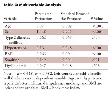
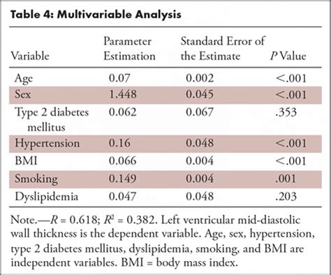
Data baserad på linjär regressionsmodell av Biaggi et al (Gender, age, and body surface area are the major determinants of ascending aorta dimensions in subjects with apparently normal echocardiograms, J Am Soc Echocardiogr. .Images used for left ventricular (LV) mid-diastolic wall thickness and LV mass measurements. (a) Prospective electrocardiographically gated CT angiography study in mid-diastolic phase of the basal, mid, and apical (from left to right) short-axis (SAX) plane with LV wall thickness caliper measurements per myocardial segment according to the .The Left Ventricle 3 1. Measurement of LV Size 3 1.1. Linear Measure-ments 3 1.2. Volumetric Measure-ments 3 1.3. Normal Reference Values for 2DE 6 . RV = Right ventricular RWT = Relative wall thickness STE = Speckle-tracking echocardiography TAPSE = Tricuspid annular plane systolic excursion TAVI = Transcatheter aortic
Fundamentals for Correct Measurement of the Left Ventricle. Some fundamental techniques to measuring the left ventricle in systole and diastole are: . The septum was measured at the tapered section of the IVS which under estimates the true thickness and severity of left ventricular hypertrophy: To diagnose left ventricular hypertrophy, a healthcare professional does a physical exam and asks questions about your symptoms and family's health history. The care professional checks your blood pressure and listens to your heart with a device called a stethoscope. Tests. Tests used to diagnose left ventricular hypertrophy may include:
With physiologic remodeling, left ventricular wall thickness rarely exceeds 15 mm and left ventricular cavity sizes tend to be larger compared with the typical left ventricular cavity sizes in HCM. 6 Diastolic function, including tissue Doppler measurements, should be normal in cases of physiologic remodeling.
Left ventricular hypertrophy (LVH) is a condition in which there is an increase in left ventricular mass, either due to an increase in wall thickness or due to left ventricular cavity enlargement, or both. Most commonly, the left ventricular wall thickening occurs in response to pressure overload, and chamber dilatation occurs in response to the volume overload.[1]

Introduction. An accurate and reproducible quantification of the left ventricular (LV) structure is important for diagnosis and monitoring of disease progression, for timing of intervention and for discrimination of prognosis. 1–3 LV chamber size and wall thickness represent the determinants of decision-making in several clinical guidelines. 1, 4, 5 .
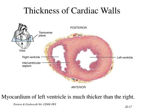
Ejection fraction (EF) is a measurement, expressed as a percentage, of how much blood the left ventricle pumps out with each contraction. An ejection fraction of 60 percent means that 60 percent of the total amount of blood in the left ventricle is pushed out with each heartbeat. A normal heart’s ejection fraction is between 55 and 70 percent.a Diagrams illustrating the three cross sections of the hearts: the first mid-ventricular one and at least two additional parallel sections towards the apex.b On the left: left ventricular (posterior), septal (middle), and right ventricular (posterior) thickness measurements, by excluding trabeculae and papillary muscles.On the right: transverse size and longitudinal size . Left ventricular mass, wall thickness, and the ratio of wall thickness to radius are important measures to characterize the spectrum of left ventricular geometry. . Fast measurement of left ventricular mass with real-time three-dimensional echocardiography: comparison with magnetic resonance imaging. Circulation. 2004; 110:1814-1818. Crossref .Average thickness of the left ventricle, with numbers given as 95% prediction interval for the short axis images at the mid-cavity level are: [8] Women: 4 – 8 mm; Men: 5 – 9 mm; CT & MRI. CT and MRI-based measurement can be used to measure the left ventricle in three dimensions and calculate left ventricular mass directly.
Table 26 Normal left ventricular myocardial thickness (in mm) in the adult measured on short axis images for men and women. Full size table. . The caliper-based linear measurement of thickness of trabeculation has progressively .
normal left ventricular wall thickness
Inspect (and measure) the left ventricular wall thickness. Valve circumferences are measured at the basal ring (bottom in image). (Measure the circumferences of the four valves. Cutoffs for valve dilatation: Mitral valve: circumference greater than 9.9 cm in males and 9.1 cm in females; Aortic valve: circumference greater than 8.5 cm in males . The ejection fraction is usually measured only in the left ventricle. The left ventricle is the heart's main pumping chamber. It pumps oxygen-rich blood up into the body's main artery, called the aorta. The blood then goes to the rest of the body. According to the American Heart Association: A left ventricle (LV) ejection fraction of about 50% .
The IVSd and IVPWd measurements are used to determine left ventricular hypertrophy, which is the thickening of the muscle of the left ventricle. LV hypertrophy is a marker for heart disease. In general, a measurement of 1.1-1.3 cm indicates mild hypertrophy, 1.4-1.6 cm indicates moderate hypertrophy, and 1.7 cm or more indicates severe hypertrophy.Assessment of left ventricular systolic function has a central role in the evaluation of cardiac disease. Accurate assessment is essential to guide management and prognosis. Numerous echocardiographic techniques are used in the assessment, each with its own advantages and disadvantages. This review is based on a literature search of the PubMed, MEDLINE, .Measurements of the left ventricle can be obtained by the MMode or by 2D methods. 3.2.2.1.1 MMode measurements / Diameter. MMode still is the most widely used the technique. The reasons are historical. . Usually you will not only measure the diameter of the ventricles but also the thickness of the septum and the posterolateral wall. In . Women with high blood pressure are more likely to get the condition than are men with similar blood pressure measurements. Complications. Left ventricular hypertrophy changes the structure of the heart and how the heart works. The thickened left ventricle becomes weak and stiff. . Left ventricular hypertrophy: Clinical findings and ECG .
Left ventricle | Radiology Reference Article | Radiopaedia.orgThe increased diastolic pressure backs up into the left atrium, pulmonary veins and pulmonary capillaries and produces congestive heart failure (CHF; i.e., pulmonary edema and pleural effusion). When LV hypertrophy is severe, it is common for the LV wall thickness to be twice the normal thickness (increased from 3 to 5 mm up to 7 to 10 mm). Measurement discrepancies of left ventricular mass and maximum wall thickness between modalities are clinically significant in patients with Fabry disease because use of one modality over the other could affect the diagnosis of left ventricular hypertrophy in 29% of patients, eligibility for disease-specific therapy in 26% of patients, and .
Left ventricular ejection fraction (LVEF) is the central measure of left ventricular systolic function. LVEF is the fraction of chamber volume ejected in systole (stroke volume) in relation to the volume of the blood in the ventricle at the end of diastole (end-diastolic volume). Stroke volume (SV) is calculated as the difference between end-diastolic volume . Cardiovascular magnetic resonance (CMR) can accurately measure left ventricular (LV) mass, and several measures related to LV wall thickness exist. We hypothesized that prognosis can be used to .

axial torsion testing atm standards
webDragon Hatch is a thrilling slot game that lets you enter the world of mythical creatures and win big prizes. Join the four elemental dragons and hatch the eggs to reveal wild symbols, free spins, and multipliers. Don't miss this exciting adventure at m.pgsoft-games.com.
left ventricle thickness measurements|left ventricular size is decreased