macular edema retinal thickness measurement|macular thickness results : custom Four techniques are examined to determine the best reference standard for the . Resultado da Aplicativo Liturgia Diária. Com o aplicativo Liturgia Diária – Canção Nova, você confere as leituras bíblicas diárias e uma reflexão do Evangelho em .
{plog:ftitle_list}
webAssista vídeos pornô de Trans Novinha de graça, aqui no Pornhub.com. Descubra a crescente coleção de vídeos e filmes Mais relevantes explícitos em alta qualidade. .
putting eye drops in pee test
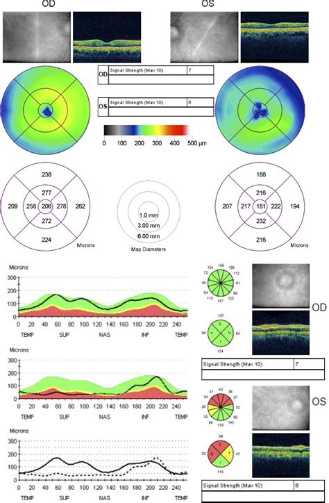
The mean and standard deviation retinal thickness by sector are shown in Figure 2 and Table 1. The foveal thickness never exceeded 252 μm in any of the healthy eyes. As expected, macular thickness was thinnest at the center, thickest within 3-mm diameter of the center, and .Four techniques are examined to determine the best reference standard for the .
Four imaging techniques that offer objective, quantitative measures of . Macular thickness measurements for a healthy eye population in this study, displayed as the mean and standard deviation in 9 regions, as . Macular thickness changes are commonly seen in eyes with retinal pathologies, such as macular thickening or edema from age-related . Optical coherence tomography (OCT) enables ophthalmologists to measure retina thickness on a cellular level and has revolutionized the way we practice ophthalmology. Considering how ubiquitous this technology is, it’s .
putting eye drops in piss test
Four techniques are examined to determine the best reference standard for the detection and quantification of macular oedema: ultrasound, optical coherence tomography, the retinal .Four imaging techniques that offer objective, quantitative measures of macular thickening are described below. Table 39 lists the axial and lateral resolutions of the four methods. TABLE . Abstract. Purpose. To determine normal macular thickness and its variation by age and gender in healthy eyes using spectral-domain optical coherence tomography (SD-OCT). Methods. Clinically, CST is an objective measurement of macular thickness readily available on OCT imaging. The central subfield is defined as the circular area 1 mm in diameter centered around the center point of the fovea, with its thickness provided as a quantifiable value on OCT software. . of retinal inner layers with visual acuity response to .
Reproducibility of Optovue RTVue Optical Coherence Tomography Retinal Thickness Measurements and Conversion to Equivalent Zeiss Stratus Metrics in Diabetic Macular Edema Transl Vis Sci Technol . 2015 Jan 22;4(1):5. doi: 10.1167/tvst.4.1.5.Volumetric analysis of early macular edema with the Heidelberg Retina Tomograph in diabetic retinopathy. Ophthalmology. 1998;105(6):1051–1059. . Schaudig UH, Glaefke C, Scholz F, Richard G. Optical coherence tomography for retinal thickness measurement in diabetic patients without clinically significant macular edema. Ophthalmic Surg Lasers .
Figure 4. A. Optical coherence tomography image of an eye with diabetic macular edema with corresponding retinal thickness mapB. generated by the OCT. C. OCT of the same patient one month following intravitreal triamcinolone acetonide injection withcorresponding retinal thickness map. D. Resolution of macular edema.Retinal thickness and retinal fluid are used to evaluate treatment response to anti-VEGF agents ().From early trials (e.g., PrONTO, PIER, and SAILOR) to more recent trials (e.g., ATLAS, TREND, and HAWK/HARRIER), retinal thickness has been used as 1 efficacy end point, 3,5,7–9,11–19 and anti-VEGF treatment has resulted in reduced retinal fluid and retinal . Diabetic macular edema (DME) is a late stage of diabetic eye disease that is characterized by retinal thickening in the macula, often accompanied by hard exudate deposition, and resultant vision loss.Traditional methods for evaluating macular edema, such as slitlamp biomicroscopy, stereoscopic photography, and fluorescein angiography, are relatively insensitive to small changes in retinal thickness and are qualitative at best. 4 The introduction of optical coherence tomography (OCT) has enabled clinicians to reliably detect and measure .
Despite these limitations, our study is the first that shows significant changes in retinal thickness measurements in diabetic macular edema after meal. The retinal thickness changes in relation to fasting state should be taken into account when evaluating the effect of a treatment modality in diabetic macular edema. To evaluate the impact of reducing B-scan frame-sampling density on retinal thickness measurements using spectral domain optical coherence tomography (SD-OCT) in eyes with diabetic macular edema .
Macular edema is a retinal condition characterized by fluid buildup in the central part of the retina responsible for sharp, central vision, and accompanies various retinal diseases such as diabetic retinopathy, retinal vascular occlusions, and uveitis.[1][2] This condition causes decreased visual acuity and, when persistent, can lead to severe vision loss.[3] Fluid .
Purpose: To compare different spectral domain optical coherence tomography devices regarding retinal thickness values in patients with diabetic macular edema and to correlate the results with conventional time domain Stratus OCT data. Methods: Thirty eyes of 30 consecutive patients with diabetic macular edema were included into a prospective study. In the short term, obstructed flow of blood and other fluids can cause acute macular edema. Over time, the starving retina will stimulate proliferation of abnormal blood vessels that can result in .The DRCR.net uses macular retinal thickness measurements to guide eligibility for participation, to apply retreatment criteria, and to serve as an outcome measure of DME in clinical trials. . Evaluation of time domain and spectral domain optical coherence tomography in the measurement of diabetic macular edema. Macular edema is the buildup of fluid in the macula, an area at the back of the eye. This fluid causes the macula to swell and thicken, which distorts vision. . NEI also funds research on diseases that cause macular edema, like diabetic retinopathy and retinal vein occlusion. Get the latest news on NEI-supported macular edema research. Last .
Compared with other measurements (versus center point thickness and total volume), it has a higher reproducibility and stronger correlation. Recent studies have shown that it is the preferred metric for . Introduction. Macular edema (ME) is one of the major causes of vision impairment in patients suffering from metabolic, vascular, and inflammatory retinal disorders (1–5).The etiology of ME includes diabetes, retinal vein .We previously have shown that OCT is an effective technique for monitoring central foveal thickness in patients with macular edema. 7. Single measurements of retinal thickness in the central fovea, however, provide an incomplete clinical picture because extrafoveal macular edema is neglected. In this study, a radial spoke pattern of six optical .
A representative cross-sectional SD-OCT scan of a patient with DM with PRVW showing retinal thickness measurement segmentation of the following layers: Inner retinal thickness—distance between . Retinal thickness is also useful for evaluating the course of disease in conditions that cause macular edema, such as diabetic macular edema, retinal vein occlusion, central serous .
Introduction. Ophthalmologists have associated diabetic macular edema (DME) and a reduction in visual acuity for decades. 1 – 4 It is well established that treatments that reduce DME can improve or stabilize visual acuity. 3, 5, 6 Using optical coherence tomography (OCT), it is now possible to measure objectively macular thickness and investigate quantitatively the . Reproducibility of spectral-domain optical coherence tomography retinal thickness measurements and conversion to equivalent time-domain metrics in diabetic macular edema. JAMA Ophthalmol. 2014; 132 (9): 1113–1122. [PMC free article] [Google Scholar]Assessment of macular thickness is important, as macular edema is a leading cause of blindness in patients with diabetes [1,2]. Clinical diagnosis of diabetic macular edema is based on the biomicroscopic examination of the fundus by observing retinal thickening and/or the presence of dry exudates and hemorrhages in the macular region.
1. Introduction. Diabetes mellitus is one of the commonest chronic diseases affecting all populations especially developed countries. Diabetic macular edema (DME), being a complication of diabetes, is an important cause of visual loss in developed countries [1, 2].Treatment of diabetic macular edema is readily available and management guidelines of . The radial traction by epimacular membranes can also result in separation of retinal layers leading to various degrees of schisis, often misinterpreted as macular edema (Figure 17). Partial-thickness macular holes are also not uncommon in the presence of epiretinal or epimacular membranes.
right eye macular thickness
Measurement of retinal thickness in MIIS-SD OCT relies on definition of the anterior and posterior retinal surfaces. This variation in macular thickness measurement seen with the Monaco OCT could be due to differences in definition of the retinal boundary, which is different in various OCT instruments (8,11). It is almost apparent that the . The correlation seen in our study between the increasing severity of retinal edema and the choroidal thickness appears to be linked to pathological mechanisms common . Berntsen D.A. Validation of macular choroidal thickness measurements from automated SD-OCT image segmentation. Optom. Vis. Sci. 2016; 93:1387. doi: . Introduction. Macular edema (ME) is one of the major causes of vision impairment in patients suffering from metabolic, vascular, and inflammatory retinal disorders (1–5).The etiology of ME includes diabetes, retinal vein occlusion (RVO), epiretinal membrane (ERM), and age-related macular degeneration (AMD) (3, 6–11).ME affects approximately 7 million people .
retinal thickness measurements
normal macular thickness study

WEBFormer Time Warner Cable and BrightHouse customers, sign in to access your roadrunner.com, rr.com, twc.com and brighthouse.com email.
macular edema retinal thickness measurement|macular thickness results