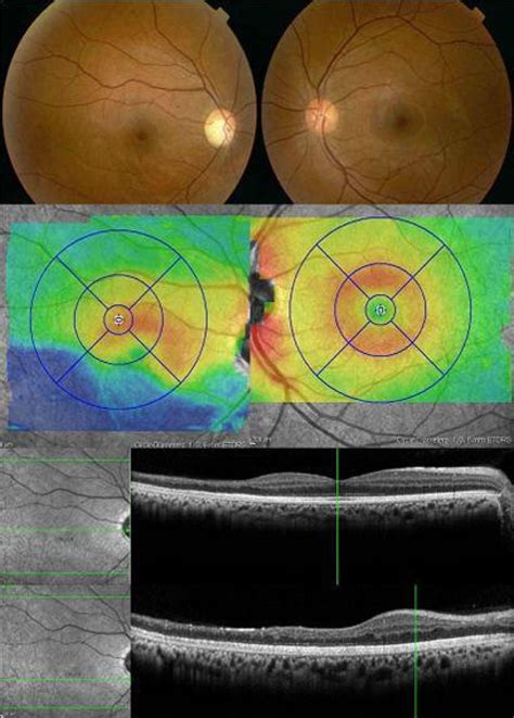
thickness measurement of retina|retinal thickness treatment : factory Recent studies have shown that it is the preferred metric for following retinal thickness measurements (i.e diabetic macular edema). 6. .
Topo de bolo Chaves. Já pensou em personalizar sua festa com um topo de bolo? Topo de bolo Chaves para imprimir são ideias econômicas para decorar doces e bolos, com apenas um papel e com palitinhos de .
{plog:ftitle_list}
John Wick 4: Baba Yaga é um filme dirigido por Chad Stahelski com Keanu Reeves, Donnie Yen. Sinopse: O assassino profissional retorna às telas .
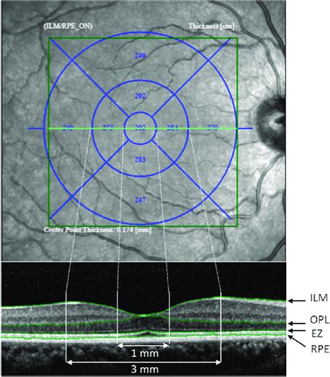
Macular thickness measurements for a healthy eye population in this study, displayed as the mean and standard deviation in 9 regions, as defined in the Early Treatment Diabetic Retinopathy Study (A) and a false-color map for a prototypical healthy eye (B).
The area of the human retina is 1,094 square mm (Bernstein, personal .An increased measurement in retinal thickness of approximately 65 to 70 .Retinal thickness and retinal fluid are common anatomical measures of .
Using spectral-domain optical coherence tomographic images (Spectralis®, wavelength: 870 nm; Heidelberg Engineering Co, Heidelberg, . Recent studies have shown that it is the preferred metric for following retinal thickness measurements (i.e diabetic macular edema). 6. .The present study measured retinal thickness with TD-OCT (Stratus) and SD-OCT (Spectralis; Heidelberg Engineering, Heidelberg, Germany) in subjects without any known retinal disease . Retinal thickness (means and standard deviations) was calculated by OCT mapping software, presented for foveal minimum, central macula .
Results. Two hundred eyes from 200 subjects (110 females, mean age 39.9±13.9 years [range 20–74 years]) were used for this study. The mean AXL was 24.30±1.07 mm (range 22.23–27.14 mm). Full retinal thickness was higher in . ThicknessTool can measure retina nuclear layers thickness in a fast, accurate, and precise manner with multi-platform capabilities. The area of the human retina is 1,094 square mm (Bernstein, personal communication), calculated from the expectations that the average dimension of the human eye is 22 mm from anterior to posterior poles, and .
32 mm from ora to ora along the horizontal meridian (Kolb, unpublished measurements). The area of the human retina is 1,094 square mm (Bernstein, . The retinal thickness shows greatest variations in the center. . Optical coherence tomography (OCT) has been applied to measure peripapillary retinal nerve fiber layer (RNFL) thickness at a micrometer scale in several optic neuropathies such as non-arteritic . Several measures of retinal thickness are used, including central retinal thickness (CRT) and central subfield thickness (CST), which measure the mean retinal thickness within the 1-mm diameter circular field surrounding the foveola, and central foveal thickness (CFT) and center point thickness, which measure the retinal thickness at the . Accurate assessment of the retinal nerve fibre layer (RNFL) is central to the diagnosis and follow-up of glaucoma. The in vivo measurement of RNFL thickness by a variety of digital imaging .
Four SD-OCT measurements of outer retinal layer thickness were selected for our analyses of outer-retinal layer related boundaries as represented in Fig. 1: inner nuclear layer -retinal pigment .
Quantitative measurements of retinal thickness and qualitative observation of retinal fluid on OCT are used as the criteria for disease activity and as efficacy outcomes in clinical trials. 3,5,7–19 In practice, clinicians evaluate OCT scans for qualitative evidence of intraretinal fluid (IRF), subretinal fluid (SRF), and irregular elevation . Retinal thickness measurements strongly correlated with those obtained by Spectralis. An increased measurement in thickness of 35.35 µm was noted in the central fovea. In addition, wide-angle fundus photography was successfully obtained in all subjects. Integrated system provides quality fundus photographs as well as OCT, obviates the need for . Measurement of retinal thickness in macular region of high myopic eyes using spectral domain OCT. Int J Ophthalmol. 7, 122–127 (2014). PubMed PubMed Central Google Scholar .
what is normal retinal thickness
Clinically, CST is an objective measurement of macular thickness readily available on OCT imaging. The central subfield is defined as the circular area 1 mm in diameter centered around the center point of the fovea, with its thickness provided as a quantifiable value on OCT software. . Here, baseline BCVA did not correlate with baseline . Optical Coherence Tomography (OCT) measurement of retinal layer thickness. Retinal layers were measured using spectral domain optical coherence tomography (SD-OCT) (Bioptigen, Research Triangle Park, NC) 2 weeks after optic nerve injury, as described . After anesthetizing the mice, the pupils were dilated and kept moist using saline.
To use Optical Coherence Tomography (OCT) to measure scleral thickness (ST) and subfoveal choroid thickness (SFCT) in patients with Branch Retinal Vein Occlusion (BRVO) and to conduct a .
The average thickness of 10 intra-retinal layers was then corrected for ocular magnification using axial length measurements, and pairwise comparisons were made using multivariable linear .Total retinal thickness measurements obtained by the algorithm were compared with measurements provided by the standard OCT3 software. Results: The total retinal thickness profile demonstrated foveal depression, corresponding to normal anatomy, with a thickness range of 160 to 291 microm. Retinal thickness measured by the algorithm and by the . We sought to compare the retinal thickness measurements collected using different optical coherence tomography (OCT) devices. This prospective study included 21 healthy cases, and the retinal thickness was measured using the PLEX Elite (Carl Zeiss Meditec, Dublin, California, USA), DRI OCT-1 Atlantis (Topcon Corp, Tokyo, Japan), Cirrus 5000 HD-OCT (Carl .
If accurate measurements of retinal layers or their thickness proportions are of interest, the automatically calculated thickness values can either be corrected by manually adjusting the delineation for the retinal pigment epithelium or by subtracting a correction factor for total retinal thickness measurements, as described in Table 3. These . Corneal thickness refers to the measurement of the cornea, which is the clear, dome-shaped surface that covers the front of the eye. . causing light to focus on multiple points instead of a single point on the retina. Corneal thickness can affect the severity of astigmatism and the success of treatments such as glasses, contact lenses, or .: An increased measurement in retinal thickness of approximately 65 to 70 μm, as measured by Spectralis OCT compared with Stratus OCT, is consistent with the extent of axial retinal thickness measured by the two instruments. This increased measurement corresponds to the inclusion of the outer segment-RPE-Bruch's membrane complex by Spectralis .
To assess retinal nerve fiber layer (RNFL) thickness measurements of normal Northern Nigerian adults using optical coherence tomography (OCT).The OCT procedure was carried out with the Carl Zeiss Stratus OCT Model 3000 software version 4.0 (Carl Zeiss . Anterior capsule thickness 14 µm Posterior capsule thickness 2–4 µm Ciliary sulcus diameter: 11.0 mm. Posterior Segment. Rod photoreceptor cells: 120 million Cone photoreceptor cells: 6 million Retinal ganglion cells (at birth): 1.2 million. 52% of optic nerve fibers cross in optic chiasm (nasal fibers) Optic Nerve Length Macular thickness changes are commonly seen in eyes with retinal pathologies, such as macular thickening or edema from age-related macular degeneration, diabetic retinopathy (DR), retinal vein occlusion, and uveitis; and macular thinning or atrophy caused by cell loss occurs in retinal dystrophies. 1–7 Evidence has shown that the degree of . Total macular thickness (internal limiting membrane to retinal pigment epithelium) is easily and accurately measured by optical reflective devices such as the OCT due to the high level of reflectivity from these two boundary regions of the retina. Earliest measurements of the retinal thickness were performed by the Retinal Thickness Analyzer .
Abstract Background: Optic disc oedema (ODE) is an important manifestation in various ocular as well as systemic disorders. Measurement of retinal nerve fibre layer (RNFL) thickness in ODE patients may help in monitoring the progress of the disease and treatment response.
retinal thickness treatment
dslreports speed test upload phase drops
Retinal thickness is a key measurement used to assess the health of the retina and whether it needs any treatment. Thickness measure of a patient’s retina should ideally fall within a range, which varies according to the region of the eye. For example, the retina is typically thinner in the center, in the foveal area. .
Purpose: Our study was conducted to establish procedures and protocols for quantitative autofluorescence (qAF) measurements in mice, and to report changes in qAF, A2E bisretinoid concentration, and outer nuclear layer (ONL) thickness in mice of different genotypes and age. Methods: Fundus autofluorescence (AF) images (55° lens, 488 nm excitation) were . Introduction The aim of this work is to investigate the differences in the measurement of foveal retinal thickness in myopic patients between two display modes (1:1 pixel and 1:1 micron) on optical coherence tomography (OCT). Methods Horizontal OCT line scan through the central fovea was used for manual measurement of foveal retinal thickness .A retinal thickness measurement is also provided. The retinal map analysis also provides a cross-sectional image and includes two maps, one with qualitative and another with quantitative thickness measurements. Measurements for nine macular sectors are shown as well as thickness measurements for the center of the scan and the total macular . Optical coherence tomography (OCT) has been found to be useful for objective measurements of retinal thickness in patients with macular edema, macular holes and epiretinal membranes with a high degree of reproducibility and repeatability [20, 21, 25].For viewing the retinal microstructure during surgery, hand-held intraoperative OCT (Intra-OP .

dslreports speed test upload phase steadily drops

retinal thickness problems
WEB19 de fev. de 2021 · Real-time Feedback. Motion capture, combined with VR training, provides employees with detailed visual feedback on how .
thickness measurement of retina|retinal thickness treatment