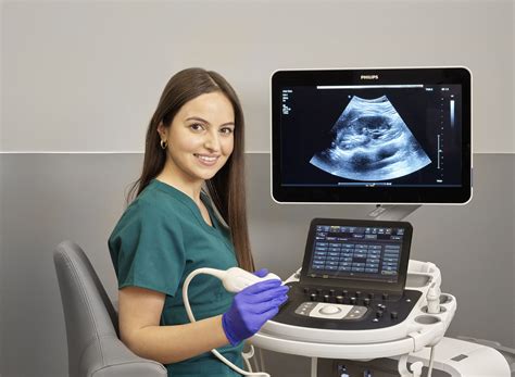
ultrasound test of soft parts|soft tissue ultrasound transducer : agencies Ultrasound is widely available, easy to use, and less expensive than most other imaging methods. Ultrasound imaging is extremely safe and does not use radiation. Ultrasound scanning gives a clear picture of soft . Deslize para a esquerda ou para a direita para comparar .
{plog:ftitle_list}
Tons of Creampie Eating porn tube videos and much more. This is the only porn resource you'll ever need!
The most common use of bedside ultrasound in patients with soft-tissue abnormalities is in the evaluation of infections, including cellulitis, abscess, and necrotizing fasciitis. Other soft-tissue indications include the evaluation of . Sonography is a diagnostic medical test that uses high-frequency sound waves, or ultrasound waves, to create images of tissues, glands, . Providers often call this test a prenatal ultrasound. Ultrasound can also check parts of your digestive system, including your: Liver. Pancreas. Gallbladder. A complete . Ultrasound is widely available, easy to use, and less expensive than most other imaging methods. Ultrasound imaging is extremely safe and does not use radiation. Ultrasound scanning gives a clear picture of soft .
Soft Tissue Neck 76536 Hypo- / hyper-thyroid E03.9/E05.90 Enlarged lymph nodes R59.9 Enlarged thyroid / fullness E04.9/E07.89 Goiter E04.9 Nodules E04.2 Palpable mass on neck R22.1 ThyroiditiE06.9 Aorta 76706 History of smoking Z87.891 Family History of AAA Z82.49 Aorta 76775 History of AAA Z82.49 AAA I71.4 Abdomen 76700What is an ultrasound? An ultrasound is an imaging test that uses sound waves to make pictures of organs, tissues, and other structures inside your body. . Ultrasound is best used to learn about conditions that involve soft tissues, .
Ultrasound images are displayed in either 2D, 3D, or 4D (which is 3D in motion). The ultrasound probe (transducer) is placed over the carotid artery (top). A color ultrasound image (bottom, left) shows blood flow (the red color in the image) in the carotid artery. Waveform image (bottom right) shows the sound of flowing blood in the carotid artery. Clinical ultrasound’s maximum utility as a diagnostic tool rests on understanding and manipulating multiple physics principles. The knowledge of ultrasound wave emission, interaction with fluid, tissue, various densities, wave receipt, and machine data processing are integral to ultrasound’s function. Ultrasound machines rely upon different probe types to emit .
what is an ultrasound

An ultrasound scan is a medical test that uses high-frequency sound waves to capture live images from the inside of your body. It’s also known as sonography.Vascular ultrasound is a noninvasive test healthcare providers use to evaluate blood flow in the arteries and veins of the arms, neck and legs. Providers use this test to diagnose blood clots and peripheral artery disease. You may also have this test to see if you’re a good candidate for angioplasty or to check blood vessel health after bypass.Ultrasound of soft parts is a diagnostic procedure used to investigate the soft tissues of the body, which include muscles, tendons, joints, and other non-bony structures. This non-invasive test employs high-frequency sound waves to create images of the internal structures, aiding physicians in diagnosing various conditions.Ultrasound, also .
Video 4. Optimizing gain; Time Gain Compensation (TGC) - Changes strength of returning echoes at various depths to help make the entire ultrasound image uniform brightness Depth Adjustment - Increases or decreases the depth of the ultrasound beam; Save - Saves an image or clip to the hard drive; Change Mode - Pushing the M-mode button will change the machine to M-mode, .
Ultrasound is sound with frequencies greater than 20 kilohertz. [1] . An ultrasonic test of a joint can identify the existence of flaws, measure their size, and identify their location. . Diagnostic ultrasound is used externally in horses for evaluation of soft tissue and tendon injuries, .Another type of ultrasound is Doppler ultrasound, sometimes called a duplex study, used to show the speed and direction of blood flow in certain pelvic organs. Unlike a standard ultrasound, some sound waves during the Doppler exam are audible. Pelvic ultrasound may be performed using one or both of 2 methods: Transabdominal (through the abdomen). However, several factors influence the resultant frequency shift and hence the measured velocity. These include the incident frequency of the ultrasound beam used, speed of sound in soft tissues, the velocity of the moving reflectors (i.e., blood in a vessel), and the angle between the incident beam and vector of blood flow (θ) called the angle of insonation.An external ultrasound scan is most often used to examine the heart or an unborn baby in the womb. It can also be used to examine the liver, kidneys and other organs in the tummy and pelvis, as well as other organs or tissues that can be assessed through the skin, such as .
Most pregnant people have an ultrasound test between 18 and 22 weeks of pregnancy. If your pregnancy is considered high-risk, . An ultrasound can diagnose health conditions that involve soft tissues like organs, blood vessels, and glands. A diagnostic ultrasound may be used to examine a growth or tumor, find a blood vessel blockage, or . Sonography is a diagnostic medical test that uses high-frequency sound waves to bounce off of structures in the body and create an image. . or ultrasound waves, to create images of tissues, glands, organs, and blood or .Find a Test; Home; Facilities; Offers; 9089 089 089. Login Home; Find A Test. Ultrasound Soft Parts. Ultrasound Soft Parts. Scans; Ultrasound; Also Known As; The scan uses high-frequency sound waves to produce images of soft tissues, such as infection, injury, and abnormal masses. Your doctor may advise this scan to evaluate infections .
An abdominal ultrasound is the most common test to screen for abdominal aortic aneurysms. Screening means looking for the condition in people without symptoms. Early diagnosis helps you and your provider take steps to manage and treat the aneurysm. If an aortic aneurysm ruptures, the bleeding can quickly lead to death. Ultrasound is a noninvasive imaging test. It uses reflected sound waves to create images or videos of soft tissues inside the body. Learn about uses and more. Ultrasound is a noninvasive imaging test. . including organs and body parts in motion. Ultrasound is safe, effective, and does not involve radiation. .Ultrasound imaging is extremely safe and does not use radiation. Ultrasound scanning gives a clear picture of soft tissues that do not show up well on x-ray images. Ultrasound is the preferred imaging modality for the diagnosis and monitoring of pregnant women and their unborn babies. Ultrasound provides real-time imaging.
For example, bones absorb more ultrasound energy than soft tissue does. Absorption of ultrasound in tissue is frequency dependent—it increases with increasing frequency. 1.8.5 Attenuation. As the ultrasound moves through tissues, some of the ultrasound energy is lost due to absorption through heat, reflection, refraction, and scattering.
An ultrasound scan is a device that uses high-frequency sound waves to create images of the inside of the body. The scans are used to assess soft tissue structures, including muscles, blood vessels, the heart and various organs. Technological advancements in the field of ultrasound now include images that can be made in a three-dimensional view (3-D) and/or four .
Sonographic artifacts from soft-tissue foreign bodies aid in their identification. Such artifacts are seen deep in relation to the foreign bodies on sonography and are not related to the composition of the material; the surface characteristics of the object influence the type of artifacts produced, particularly “clean” versus “dirty” shadowing [].1. Preparation : Preparation for a small parts ultrasound depends on the specific area to be examined.Generally, no special preparation is needed unless otherwise instructed by your doctor. 2. Clothing : It is advised to wear loose, comfortable clothing that can be easily moved or removed to expose the area being examined.You may be provided with a hospital gown if necessary. It can help diagnose problems with soft tissues, muscles, blood vessels, tendons, and joints. . A doctor or a specially-trained sonographer will carry out the test. External ultrasound.
Contact ultrasonic inspection can be performed where only one side of a test specimen as reachable, or where the parts to be tested are large, irregular in shape or difficult to transport. Immersion ultrasonic testing is a laboratory-based or factory-based non-destructive test that is best suited to curved components, complex geometries and for . The Society of Radiologists in Ultrasound convened a panel of specialists from radiology, orthopedic surgery, and pathology to arrive at a consensus regarding the management of superficial soft-tissue masses imaged with US. The recommendations in this statement are based on analysis of current literature and common practice strategies. This statement .
ultrasound for soft tissue aspiration
Sarcoma is a type of cancer that starts in certain parts of the body, like bone or muscle. These cancers start in soft tissues like fat, muscle, nerves, fibrous tissues, blood vessels, or deep skin tissues. They can be found anywhere in the body, but most of them start in the arms or legs. . Ultrasound: This test uses sound waves to make .

ford 6.7 compression tester
ultrasound for soft tissue
TV-MA. Genre. Drama. Original Language. English. Release Date. Jun 4, 2023. After a nervous breakdown derailed Jocelyn’s last tour, she’s determined to claim her rightful .
ultrasound test of soft parts|soft tissue ultrasound transducer