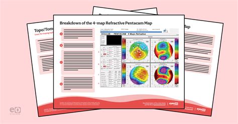
measure anterior posterior thickness|Left Ventricular Mid : chain store Numerical overlays will confirm steepening and thinning and demonstrate central corneal thickness, maximum keratometry, and anterior and posterior corneal elevations. web29 de jul. de 2019 · A Versão Atual É 9.2, Atualizada Em 29/07/2019 . De Acordo Com O Google Play, O NovelasFlix 2.0 Alcançou Mais De Instalações 68 Mil. Atualmente, O .
{plog:ftitle_list}
WEB3 dias atrás · The Good Doctor season 7 next episode. Another all new episode of The Good Doctor season 7 airs on Tuesday, February 27, at 10 pm ET/PT on ABC. The latest episode is titled "Skin in the Game," here is the official synopsis: "Shaun struggles to accommodate the newest member of his surgical team, Charlie, who interferes in a .
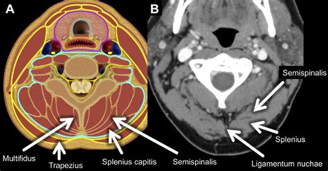
Perivertebral space
seed food grain digital moisture meter
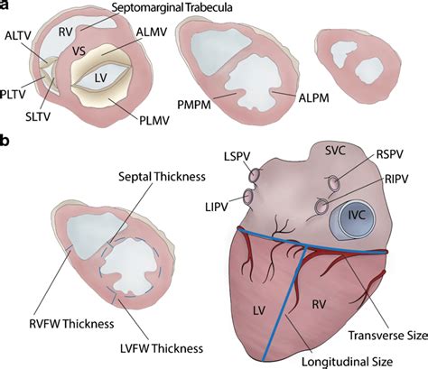
Left Ventricular Mid
The perivertebral space is a cylinder of soft tissue lying posterior to the retropharyngeal space and danger space surrounded by the prevertebral layer of the deep cervical fasciaand extends from the skull base to the upper mediastinum. The deep cervical fascia sends a deep slip to the transverse process . See moreIn general, the prevertebral componentis measured on sagittal imaging as the distance between the anterior border of the vertebral body . See more This study found that the LV was thickest in the basal septum (segment 3) with a mean thickness of 8.3 mm and 7.2 mm and thinnest in the .
Numerical overlays will confirm steepening and thinning and demonstrate central corneal thickness, maximum keratometry, and anterior and posterior corneal elevations. In adults, the normal kidney is 10-14 cm long in males and 9-13 cm long in females, 3-5 cm wide, 3 cm in antero-posterior thickness and weighs 150-260 g. The left kidney is usually slightly larger than the right. . The kidney is bean-shaped with a superior and an inferior pole, anterior and posterior surfaces, and lateral and medial borders. A full-thickness rotator cuff tear is characterized by a focal transmural tendon discontinuity, . Indirect signs on MRI are - subdeltoid bursal effusion, particularly if anterior, medial dislocation of biceps, fluid along biceps tendon and diffuse loss of peribursal fat planes.

Corneal imaging is widely used by ophthalmologists to understand the shape and curvature of the cornea. Corneal topography evaluates the anterior surface of the cornea and displays the information using a color-coded map. On the other hand, corneal tomography takes into account the thickness of the cornea, allowing the posterior surface of the cornea to be . Endometrial thickness is a commonly measured parameter on routine gynecological ultrasound and MRI. The appearance, as well as the thickness of the endometrium, will depend on whether the patient is of reproductive age or postmenopausal and, if of reproductive age, at what point in the menstrual cycle they are examined. . The .
unimeter grain moisture meter
LV Volume Measurement 2D Method s LVID Diastole (LVIDD) Inner edge to inner edge, perpendicular to the long axis of the LV, . RWT= (2x posterior wall thickness)/(LVIDD). Useful to categorize LV mass and pattern of remodeling. 4 Left Ventricular Function Assessment Purpose To measure peripapillary retinal nerve fiber layer (RNFL) thickness and posterior pole retinal thickness in primary angle-closure suspects (PACS) by Spectral domain optical coherence tomography (SD-OCT) and to be compared with normal subjects. Methods Thirty five primary angle-closure suspect patients and thirty normal subjects were enrolled in . Lesion classification is of high clinical importance in determining the right treatment option. While the Outerbridge Classification, published in 1961, takes the lesion size into consideration (Grade 2: partial thickness lesions 〈1.5 cm in diameter; Grade 3: lesions 〉1.5 cm in diameter or full thickness), the widely used International Cartilage Repair Society .Endometrial thickness is measured as the maximum anterior–posterior thickness of the endometrial echo on a long-axis transvaginal view of the uterus. The earliest reports comparing transvaginal ultrasonography with endometrial sampling consistently found that an endometrial thickness of 4–5 mm or less in women with postmenopausal bleeding .

Urinary bladder wall thickening - Radiopaedia.org the anterior cuff (subscapularis) functions to balance the posterior moment created by the posterior cuff (infraspinatus and teres minor) this maintains a stable fulcrum for glenohumeral motion. the goal of treatment in rotator cuff tears is . We then focus on the flow divider (the junction between the internal and the external carotid artery) from an anterior ( Fig. 6.8B), lateral ( Fig. 6.8C), and posterior projection ( Fig. 6.8D), finding the one projection that best displays the full extent of the plaque ( Fig. 6.8B). The probe can be placed lower or higher in the neck depending .
A Single-line scan passing through the temporal scleral reflex.B Cropped raw B-scan image of the anterior sclera (dimension of the exported image: length: 9 mm, depth: 2.6 mm).The yellow arrowheads indicate the anterior scleral boundary and posterior scleral boundary, and the red arrowhead indicates the location of the scleral spur (reference point).
Once the anterior and posterior surfaces of the kidney have been fully imaged in the sagittal plane, the transducer is rotated to obtain a transverse image. . the medullary pyramids are often indistinct on ultrasound imaging making the measurement of cortical thickness inaccurate. Therefore, parenchymal thickness is often easier to measure. A .
Corneal indices were performed with two modalities in both eyes including; apical corneal thickness (ACT), corneal thickness at pupil site(PCT), thinnest corneal thickness (TCT), anterior chamber .
A, This anterior placenta measured 7.1 cm in thickness at 28 weeks’ gestation. The fetus was hydropic in the setting of trisomy 21. B, The placental thickness measures 4.6 cm at 20 weeks’ gestation. .
CBCT can be considered a relatively reliable method for gingival thickness measurement in both the anterior and posterior areas compared with direct probing. Ultrasonic devices provide limited accuracy in the . The ratio between the anterior and posterior wall thickness is calculated. A ratio of around 1 indicates that the myometrial walls are symmetrical and a ratio well above or below 1 indicates asymmetry, although this may also be estimated subjectively (Figure S3). . From where the measurement(s) should be taken to calculate this ratio depends . The AUC values for ultrasonographic measurement in the anterior and posterior regions were 0.681 and 0.597, respectively. While the sensitivity values for measurement of the gingival thickness in the anterior and posterior regions were equal (0.833), the specificity of measurement in the anterior region (0.611) was slightly higher than the .Posterior corneal surface contributes approximately 0.4 D of against-the-rule astigmatism. . Capsule thickness: Anterior capsule: 14.0-15.5 microns; Thinnest point: Posterior capsule: . Zhou C, Jiang C, Liu G, Sun X. Measurement of Iris Thickness at Different Regions in Healthy Chinese Adults. J Ophthalmol. 2021 May 11;2021:2653564. doi: 10 .
A variety of factors guide the evaluation and management of burns. First is the type of burn, such as thermal, chemical, electrical, or radiation. Second is the extent of the burn, usually expressed as the percentage of total body surface area (%TBSA) involved. Next is the depth of the burn described as superficial (first degree), partial (second degree) or full .
Endometrial thickness is measured perpendicular to the long axis of the uterus in a midsagittal plane, including both the anterior and posterior endometrial lining, excluding the hypoechoic sub-endometrial zone. In the case of fluid collection, it . In Placido-disc systems only the anterior surface is measured; Scheimpflug systems measure both anterior and posterior elevations. 1-4; . the reference slit beam surface and the reflected beam captured by the camera can be used to analyze the anterior and posterior curvature and corneal thickness. The Orbscan (Bausch+Lomb, Rochester NY) . In another study, Baumgaertel 12 used 30 dry skulls to assess bone thickness in the coronal sections and used dental contacts as a landmark to measure sections in the anterior–posterior dimension. He reported that bone thickness in the anterior is greatest and gradually decreases posteriorly.
The average anterior wall thickness was 1.1 ± 0.2 mm; posterior wall thickness was 1.1 ± 0.2 mm, luminal diameter 2.2 ± 0.6 mm, and external elastic membrane (EEM) diameter 4.5 ± 0.9 mm. The bias of the measurements within the same operator for LAD wall thickness, luminal diameter, and EEM was 0.042, -0.06, and -0.077 mm, respectively. Devices now are able to characterize both the anterior and posterior corneal surfaces, creating a three dimensional map. . Axial maps are less sensitive at measuring the corneal curvature and, thus, are used mainly for screening purposes (4-5). . The central corneal thickness is estimated to be 525µm, and the residual stromal bed is .
Average thickness was measured in each region, giving 60 thickness measurements/eye. Scleral geometric features were correlated with globe axial length. Results: : Group mean thickness over the whole sclera was 670±80 µm (mean±SD). Maximum thickness occurred at the posterior pole of the eye, with mean thickness of 996±181 µm.
A supraspinatus tear is a tear or rupture of the tendon of the supraspinatus muscle. The supraspinatus is part of the rotator cuff of the shoulder. Most of the time, it is accompanied by another rotator cuff muscle tear.This can occur due to trauma or repeated micro-trauma and present as a partial or full-thickness tear. Quite often, the tear occurs in the tendon or as an .

Fake Insta - Fake Chat And Pos WhatsFake - FakeChat · Entretenimento. Tv Aberta Lite Flavia Devs · Entretenimento. Himawari Uzumaki Next Generation Wallpaper HD hashirama · Entretenimento. AppTV - Live Global TV channel Extol Ventures · Entretenimento. C-PRO i9Soluções · Entretenimento. Messi Psg Wallpaper ANDROID_WORLD · .
measure anterior posterior thickness|Left Ventricular Mid