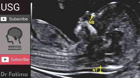
ultrasound fetal nuchal thickness measurement|ultrasound for nuchal translucentness : store The nuchal fold is a normal fold of skin seen at the back of the fetal neck during the second trimester of pregnancy. Increased thickness of the nuchal fold is a soft marker associated with multiple fetal anomalies, and is measured on a routine second trimester . web12 de set. de 2022 · Mi tío. Andy intenta regresar con su novia Mía y convence a Tadeo para que lo ayude a formar una banda de rock. Tadeo acepta y a cambio le pide ayuda para .
{plog:ftitle_list}
Old, single-lined slots had no reason to give false wins, wher.
The nuchal fold is a normal fold of skin seen at the back of the fetal neck during the second trimester of pregnancy. Increased thickness of the nuchal fold is a soft marker associated with multiple fetal anomalies, and is measured on a routine second trimester .
Thickness of the translucency varies with gestational age: Peak thickness at 12-13 weeks (in 75% of fetuses). At 12-13 weeks the 50th percentile thickness = 1.7mm. At 12-13 weeks the 95th percentile thickness = 2.8mm. Most authors .Measurements of fetal nuchal translucency, nasal bone and facial angle according to the standards of the Fetal Medicine Foundation . an "average" nuchal thickness of 2.18mm has been observed; however, up to 13% of chromosomally normal fetuses present with a nuchal translucency of greater than 2.5mm. . study values of 79.6% and 2.7% for the .Some authors recommend measuring the nuchal thickness twice and averaging the values (1), while others advocate measuring it three times and using the largest value for risk assessment (2). Measure between 11 weeks and 14 .
A system for the automatic measurement of the nuchal translucency thickness from ultrasound video stream of the foetus. In Proceedings of the 26th IEEE International Symposium on Computer-Based .
Background: The first-trimester ultrasound assessment of nuchal translucency (NT) thickness has lately been recommended as the most helpful sign in early screening for prenatal chromosomal disorders. Increased foetal NT thickness between 11 and 13+6 weeks of gestation is a frequent phenotypic manifestation of chromosomal abnormalities as well as a .The amount of fluid is measured during a nuchal translucency (NT) ultrasound scan, usually during your dating scan at around 12 weeks (PHE 2021a, PHS 2023a, PHW 2022). . Do early fetal measurements and nuchal translucency correlate with term birth weight? J Obstet Gynaecol Can 39(9):750-756.Nuchal fold measurement is obtained from the outer edge of the occipital bone to the skin surface in the transaxial plane of the fetal head at the level of the cavum septi pellucidum and cerebellar hemisphere. . , Fassnacht MA et.al. Routine measurement of nuchal thickness in the second trimester. J Matern Fetal Med 1992; 1:82-86 ;

Nuchal Translucency Thickness Measurement in Fetal Ultrasound Images to Analyze Down Syndrome. . The most salient pre-natal marker to identify DS during the early stages of gestation is the Nuchal translucency (NT) thickness. Accurate NT measurement from ultrasound (US) images becomes challenging due to the presence of speckle noise, weak . What is the nuchal translucency test? The nuchal translucency test (also called the NT scan) uses ultrasound to assess your developing baby's risk of having Down syndrome (DS) and some other chromosomal abnormalities, as well as major congenital heart problems. It's offered to all pregnant women, along with a blood test, in first-trimester .
Nuchal Translucency Normal Range Chart. When the nuchal scan is done, the doctor will share the results with you. At that time, it is important to understand what a normal measurement is. For a baby that is between 45 mm and 84 mm in size, a normal measurement is anything less than 3.5 mm. The NT grows in proportion to the baby.
Increased fetal NT thickness refers to the measurement of the vertical thickness in the midsagittal section of the fetus that is equal to or greater than a specific threshold. . the incidence of aneuploidy by nuchal thickness measurement was 7% with an NT between the 95th percentile for crown-rump length and 3.4 mm, 20% with NT between 3.5 mm .The screening is a blood test that evaluates substances in the blood (analytes), and NT is a sonogram that looks at nuchal translucency in the back of the fetal neck. Noninvasive perinatal testing (NIPT) is a newer method that provides a result with a blood test only; a first trimester ultrasound is still recommended.DOI: 10.1080/03772063.2021.1972847 Corpus ID: 240520960; Nuchal Translucency Thickness Measurement in Fetal Ultrasound Images to Analyze Down Syndrome @article{Thomas2021NuchalTT, title={Nuchal Translucency Thickness Measurement in Fetal Ultrasound Images to Analyze Down Syndrome}, author={Mary Christeena Thomas and . While both measurements are at the level of the fetal head or neck, a nuchal fold thickness, which is only performed in the second trimester, should not be confused with a first trimester nuchal translucency (NT) measurement. If an enlarged second trimester nuchal fold measurement is obtained, next steps should include. Detailed anatomic study
The nuchal translucency (NT) is an ultrasound measurement defined as the collection of fluid under the skin behind the neck of the fetus obtained between 10 and 14 weeks’ gestation (crown–rump length between 38–45 and 84 mm) (Fig. 12.1).While some fluid is present in the nuchal space of all fetuses, regardless of chromosomal status, it tends to increase among .
ultrasound for nuchal translucentness
An NT scan, or nuchal translucency scan, is a non-invasive ultrasound screening for Down syndrome and other genetic conditions during pregnancy. It’s usually done between weeks 11 and 14 of .
DOI: 10.1117/12.2075288 Corpus ID: 122528529; Ultrasound semi-automated measurement of fetal nuchal translucency thickness based on principal direction estimation @inproceedings{Yoon2015UltrasoundSM, title={Ultrasound semi-automated measurement of fetal nuchal translucency thickness based on principal direction estimation}, author={Hee . The measurement of nuchal translucency (NT) thickness in ultrasound images of the fetus at 11–14 weeks' gestation is used, in conjunction with maternal age, as an indicator of the risk of chromosomal defects 1, 2. Nuchal translucency thickness is measured manually by placing the ultrasound scanner cursors on the edges of the translucency . Nuchal cord can have clinical implications on NT and nuchal fold thickness (NFT) measurements, in the first and second trimesters of the pregnancy, respectively . . Taipale et al., have shown that there is a learning curve in ultrasound detection of fetal anomalies in early pregnancies at 13 to 14 weeks. Although the detection rate was only .
What is a nuchal translucency scan? A nuchal translucency scan (also called an NT or nuchal scan) uses ultrasound to assess your baby’s risk of having Down syndrome and some other chromosomal abnormalities, as well as major congenital heart problems. You may be offered a nuchal scan as part of your prenatal screening (Audibert et al 2017, Chitayat et al 2017, .
The nuchal translucency test measures the nuchal fold thickness. This is an area of tissue at the back of an unborn baby's neck. . Your health care provider uses abdominal ultrasound or a vaginal ultrasound to measure the nuchal fold. All unborn babies have some fluid at the back of their neck. . Nuchal translucency. In: Copel JA, D'Alton . Comparison of nuchal skin fold thickness (NFT) in a normal 20-week fetus in the breech and transverse presentations. (a) A sonogram in the breech presentation demonstrates an increased NFT measurement of 6.2 mm. The fetal head .Objective: To estimate intersonographer and intrasonographer variance components of fetal nuchal translucency (NT) thickness measurement using the traditional manual approach and a new semi-automated system. Methods: A semi-automated method was developed for measurement of the NT. In this method, the operator places an adjustable box over the . Objective To investigate a new method of screening for fetal trisomies on the basis of maternal age and fetal nuchal translucency thickness at 10 to 13 weeks of gestation.Design A prospective .
Ultrasound image based fully-automated nuchal translucency segmentation and thickness measurement January 2021 The International Journal of Nonlinear Analysis and Applications (IJNAA) 12(Special .The nuchal translucency test measures the nuchal fold thickness. This is an area of tissue at the back of an unborn baby's neck. . problems in the baby. How the Test is Performed. Your health care provider uses abdominal ultrasound or a vaginal ultrasound to measure the nuchal fold. All unborn babies have some fluid at the back of their neck .The ultrasound clinic will send a full report to your doctor. The full results may take 7 to 10 days. What do the nuchal translucency scan results mean? Your nuchal translucency scan measurement is combined with a blood test to work out the ‘combined first-trimester screen’ result. The calculation also includes your age.
The objective of the paper is to introduce a novel method for nuchal translucency (NT) boundary detection and thickness measurement, which is one of the most significant markers in the early screening of chromosomal defects, namely Down syndrome. To improve the reliability and reproducibility of NT measurements, several automated methods have been .
nuchal translucent thickness chart
WEBMathematics, Computer Science and Natural Sciences Faculty 1. Architecture Faculty 2. Civil Engineering Faculty 3. Mechanical Engineering Faculty 4. Georesources and Materials Engineering Faculty 5.
ultrasound fetal nuchal thickness measurement|ultrasound for nuchal translucentness