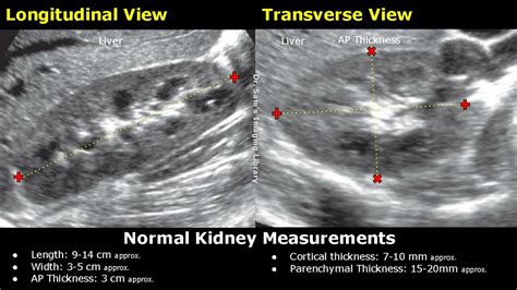
ultrasounds measure parenchymal thickness kidneys|kidney ultrasound results chart : trading The kidneys are located to either side of the vertebral column in the perirenal space of the retroperitoneum, within the posterior abdominal wall. The long axis of the kidney is parallel to the lateral border of the psoas muscle and it lies anterior to the quadratus lumborum muscle. Being parallel to the psoas muscle, . See more Resultado da 19 de mai. de 2023 · With Mortal Kombat 1 having finally been announced, 2023 officially becomes one of the best years on record for the fighting games esports scene. We’re mere days away from the launch of Street Fighter 6, and on September 19th, we’ll get access to Mortal Kombat 1. This year, .
{plog:ftitle_list}
webFounded 1903 Address Parque de la Independencia 2000 Rosario, Santa Fe Country Argentina Phone +54 (341) 411 7725 Fax +54 (341) 411 7729 E-mail [email protected]
torsion testing machine manual
The kidneys are located to either side of the vertebral column in the perirenal space of the retroperitoneum, within the posterior abdominal wall. The long axis of the kidney is parallel to the lateral border of the psoas muscle and it lies anterior to the quadratus lumborum muscle. Being parallel to the psoas muscle, . See moreA discrepancy of >2 cm between renal lengths should be considered abnormal 10and may indicate an underlying disease. Common diseases affecting the kidneys include: 1. gamuts 1.1. delayed nephrogram 1.2. persistent nephrogram 1.3. striated . See more
Kidney length should not be less than three vertebral body lengths, and no more than four vertebral body lengths 10. Antenatally, fetal . See moreThe collecting system arises from the ureteric bud, which arises from the mesonephric duct in the fourth week of gestation. The renal parenchyma arises from the metanephros, which appears in the fifth week, a derivative of the intermediate . See more The parenchymal thickness is measured from the renal capsule to the edge of the renal sinus. In adults, the medullary pyramids are often indistinct on ultrasound imaging .The kidneys are easily examined, and most pathological changes in the kidneys are distinguishable with ultrasound. In this pictorial review, the most common findings in renal .
torsion testing machine manufacturers in india
Our study attempted to evaluate the usefulness of a generally obtainable measurement at ultrasound in the setting of CKD as a correlate to kidney . OBJECTIVE. The objective of our study was to develop, by use of ultrasound, nomo-grams of renal parenchymal thickness, medullary pyramid thickness (height), renal . Ultrasound imaging is a key investigatory step in the evaluation of chronic kidney disease and kidney transplantation. It uses nonionizing radiation, is noninvasive, and generates real-time images, making it the ideal initial .Sample image of shear wave elastography used to assess a kidney’s parenchymal stiffness. (A) A confidence map is used to obtain high-quality measurements. (B) The elastogram .
Increased length with a rounded appearance and loss of sinus fat is indicative of parenchymal swelling, whereas a small, lobulated kidney suggests cortical thinning. The thickness of the cortex should be determined when possible, . Measure cortical thickness: Cortical thickness should be estimated from the base of the pyramid and is generally 7–10 mm. If the pyramids are difficult to differentiate, the parenchymal thickness can be measured instead .
The cortical thickness is measured from the base of the pyramid to the capsule and is normally between 6 and 10 mm. If the pyramids are difficult to see, the parenchymal thickness can be measured, instead of measuring . The accuracy of US measurement was further assessed using Bland–Altman plots for agreement between US and MRI measurements for length, volume, and parenchyma thickness of right and left kidneys . .What is the value of measuring renal parenchymal thickness before renal biopsy? Clin Radiol. 1994; 49:45-49. Google Scholar. 20. . Ultrasound assessment of liver and kidney brightness in infants: Use of the gray-level . The kidneys measure about 5 to 7 cm in width, 3 to 5 cm in thickness, . the parenchymal thickness can be measured, . Some ultrasound (US) units can calculate the renal volume from the three .
Like renal length, parenchymal thickness gives an indication of the chronicity of renal failure. However, some patients with parenchymal thickness 1.5 cm or less still have potential for improvement. This measurement alone should not be used to obviate renal biopsy. Ultrasonography of the kidneys is essential in the diagnosis and management of kidney-related diseases. The kidneys are easily examined, and most pathological changes in the kidneys are distinguishable with ultrasound. In this pictorial review, the most common findings in renal ultrasound are highlighted.
Renal parenchymal disease is a group of conditions that can develop in the parenchyma of your kidneys. The parenchyma is the functional part of an organ, as opposed to the parts that give the .
Objective: The objective of our study was to develop, by use of ultrasound, nomograms of renal parenchymal thickness, medullary pyramid thickness (height), renal length, and the ratio of medullary pyramid thickness to parenchymal thickness in healthy children. Subjects and methods: This prospective study included 292 consecutive children (136 boys and 156 girls) . Then, the predicted masks were used to estimate the kidney image biomarkers, including its volume and dimensions (length, width, thickness, and parenchymal thickness). Normal child kidney ultrasound scan image: *renal column, **renal pyramid, ***renal cortex, ****renal sinus . the thickness of the renal parenchyma can be measured and is 15-20 mm. Renal measurements: L=length; P=parenchymal thickness; C=cortical thickness. . Hydronephrosis can also be assessed by measuring the level of the neck in the . Methods such as measuring the parenchymal thickness, RV to parenchymal thickness ratio, renal artery Dopplers and echogenicity of the renal parenchyma are potential non-invasive methods to evaluate nephron endowment and future renal function. . Evaluation of fetal kidney growth using ultrasound: a systematic review. Eur J Radiol 2017; 96:55 .
An adverse intrauterine environment may result in a depleted nephron number and an increased risk of chronic kidney disease. Antenatal ultrasound was used to measure the foetal renal parenchymal thickness (RPT), as a novel method to estimate nephron number. Foetal renal artery blood flow was also assessed.This is a unitless, surrogate measure of intrarenal parenchymal pathology, defined as the ratio of the difference between peak systolic velocity and end-diastolic velocity to the peak systolic velocity. . They reported a correlation between RRI and increased common carotid thickness but not kidney-specific . Gutteridge C.M. The clinical use .In growth restricted fetuses the renal parenchymal thickness was found to be significantly less when compared to the parenchymal thickness of appropriately grown fetuses. Conclusions: Measurement of the renal parenchymal thickness is an innovative method to evaluate the development of the fetal kidneys.
When possible, these measurements were made on the kidney that was included in the study. Measurements at three different areas were made in each examination, with sufficient magnification, and then the average of these measurements was used. Fig. 1. A representative longitudinal sonogram for the measurement of renal cortical thickness.The aim of this study was to obtain ultrasound measurements of PT in children with normal SFK and to compare these data with PT measurements in children with two functioning kidneys. Methods: This was a prospective multicenter study carried out between 2006 and 2011 in which 236 children aged 11 days to 18.96 years with healthy SFK were examined.
Renal parenchyma thickness measurement using ultrasonography is a novel non-invasive measurement of renal . A kidney ultrasound examination was performed at 32- and 37-weeks postmenstr- .OBJECTIVE. The objective of our study was to develop, by use of ultrasound, nomograms of renal parenchymal thickness, medullary pyramid thickness (height), renal length, and the ratio of medullary pyramid thickness to parenchymal thickness in healthy children. SUBJECTS AND METHODS. This prospective study included 292 consecutive children (136 boys and 156 girls) . Renal parenchyma thickness measurement using ultrasonography is a novel non-invasive measurement of renal development for the determination of nephron endowment. . The kidney ultrasound .
Ultrasonographic kidney measurements were performed on 66 adult patients with known diabetes and hypertension with different duration of the disease. The impact of age, gender, site (left and right side) and height was statistically analyzed. . Renal length and parenchymal thickness were assessed by ultrasound. The study was carried out .Previous studies have shown that renal volume calculated at ultrasound is a more exact measurement of a functioning kidney than renal length [2, 3]. . They also showed the addition of renal area and parenchymal thickness measured at ultrasound to length was a better predictor of both single-kidney GFR and renal volume than length measured at .
ultrasound for right renal unit
Multiple renal measurements were performed on both kidneys at each scan. The renal parenchymal thickness was measured in the mid-sagittal plane. Standard charts were developed and the intra and interobserver reliability for the renal measurements was analysed. Results. Standard charts were developed for fetal renal parenchymal thickness, length . Patients were positioned in the left lateral decubitus position for two-dimensional ultrasound examinations of the right kidney. The examinations included observing kidney morphology and parenchymal echo, and measuring the kidney’s thickness, diameter, parenchyma thickness, and the skin-to-capsule distance. Causes of echogenic kidneys on ultrasound can include: acute and chronic kidney disease, sickle cell disease, kidney disease related to HIV, renal amyloidosis, and dehydration. Is echogenic kidney dangerous? It can be in some cases when the cause is related to kidney disease. More testing will be needed to be certain.
The posterior parenchymal thickness was used as it was closest to the ultrasound transducer and our previous study demonstrated no significant difference between the anterior and posterior thickness. 20 For each kidney, the measurement was performed twice and the mean of the two measurements was recorded. Length of 9-12 cm and width of 4-5 cm in the normal adult. The left kidney is often slightly larger than the right, but both should be within 2 cm of each other. Figure 1. Measurement of normal right kidney in longitudinal view; There are many normal variations in the anatomic structure of the kidneys. Some common ones that you may identify .
Methods: Renal ultrasound (US) parameters including renal length, parenchymal thickness, cortical thickness and medullary thickness were assessed in 176 subjects, who were categorized into 4 groups based on the estimated glomerular filtration rate (ml/min/1.73 m 2): group 1, ≥90; group 2, ≥60 but <90; group 3, ≥30 but <60; and group 4 .An adverse intrauterine environment may result in a depleted nephron number and an increased risk of chronic kidney disease. Antenatal ultrasound was used to measure the foetal renal parenchymal thickness (RPT), as a novel method to estimate nephron number. Foetal renal artery blood flow was also assessed.
ultrasound for kidney volume
renal ultrasound parenchymal thickness

WEBLiveTV is a free website for live sport streams, sport videos and live score. We offer you a great possibility to follow numerous live sport events. LiveTV helps fans all over the world to watch live US football , basketball, soccer, hockey and many other sports. Right from the stadium to your living room. All the sport streams are free, we don .
ultrasounds measure parenchymal thickness kidneys|kidney ultrasound results chart