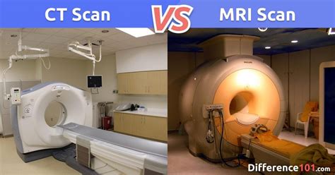
what test will allow you to see soft tissue|CT Scan Versus MRI Versus X : factories X-ray imaging is often appropriate for detecting issues with the bones, while an MRI might be more appropriate if a doctor suspects any issues with your body’s soft tissues. Whether you need an x-ray, CT, DEXA, MRI or ultrasound, . WEB1 - 1. ontem. Campeonato Espanhol. Real Betis. Deportivo Alavés. 0 - 0. DOM, 18/02. Liga Conferência Europa da UEFA. Real Betis. Dinamo Zagreb. 0 - 1. QUI, 15/02. .
{plog:ftitle_list}
1 de fev. de 2024 · O Betsat é um aplicativo que veio para transformar a forma como os fãs interagem com o seu esporte favorito, oferecendo uma experiência única e .
MRI may be used to assess the results of corrective orthopedic procedures. Joint deterioration resulting from arthritis may be monitored by using magnetic resonance imaging. There may be other reasons for your physician to .
Less dense soft tissues and breaks in bone let radiation pass through, making these parts look darker on the X-ray film. You will probably be X-rayed from several angles. If you have a fracture in one limb, your doctor may want a .The soft tissues in the body (such as blood, skin, fat, and muscle) allow most of the X-ray to pass through and appear dark gray on the film or digital media. A bone or a tumor, which is more dense than soft tissue, allows few of the X .X-ray imaging is often appropriate for detecting issues with the bones, while an MRI might be more appropriate if a doctor suspects any issues with your body’s soft tissues. Whether you need an x-ray, CT, DEXA, MRI or ultrasound, . Early testing in pig tissue has shown that STAR can remove tumours and cut tissue as precisely as surgeons can — a crucial skill because leaving even a few tumour cells behind could allow the .
Radiography is the imaging method which uses x-rays or electromagnetic waves.These waves pass through the person’s body, with some rays being absorbed by the tissues and others reaching the radiographic film behind. Thus creating a 2 dimensional (flat) image called a radiograph.Dense tissues will absorb most of the rays and come out on the .
Magnetic Resonance Imaging (MRI) of the Bones,
CT Scan Versus MRI Versus X
MRI, which uses powerful magnets to produce 3-D anatomic images, is a high-contrast resolution modality that can determine changes in the tissue quality.Study with Quizlet and memorize flashcards containing terms like 1. When assessing a patient, consider the possibility of closed soft-tissue injuries whenever there is swelling, pain, or deformity, as well as: A. a medical condition that would explain this presentation. B. a mechanism of blunt trauma. C. signs of underlying fractures. D. penetrating trauma., 2. An internal injury . Treatment for soft tissue injuries depends on several factors, including:. the severity; the type of injury; the particular joint, muscle, or limb affected; People can often self-treat mild soft . CT images of internal organs, bones, soft tissue, and blood vessels provide greater detail than traditional x-rays. This is especially true for soft tissues and blood vessels. Using specialized equipment and expertise to create and interpret CT scans of the body, radiologists can more easily diagnose problems such as cancer, cardiovascular .
You may need a follow-up exam. If so, your doctor will explain why. Sometimes a follow-up exam further evaluates a potential issue with more views or a special imaging technique. It may also see if there has been any change in an issue over time. Follow-up exams are often the best way to see if treatment is working or if a problem needs attention. Soft tissues (such as muscle and body organs) show up as various shades of grey, depending on how dense they are. The developed film is studied by an X-ray doctor (radiologist) who sends a report to the doctor who requested the test. An ordinary X-ray test is painless. You cannot see or feel X-rays. Some X-rays use contrast material (also called contrast agent or dye). It makes certain structures in your body, like blood vessels, easier to see. The contrast material comes as a liquid, powder or pill. Your provider gives you the contrast material before the X-ray. Depending on the type of X-ray, you may receive the contrast material:
 of the Bones, .jpg)
Tests show I have a benign soft tissue tumor. Should I be worried? Remember, a benign soft tissue tumor isn’t cancer. Benign soft tissue tumors are about 10 times more common than malignant or cancerous soft tissue tumors. In general, you could have cause for concern if a bump or lump that’s a soft tissue tumor affects your quality of life .Reticular tissue is a mesh-like, supportive framework for soft organs such as lymphatic tissue, the spleen, and the liver (Figure 6). Reticular cells produce the reticular fibers that form the network to which other cells attach. It derives its name from the Latin reticulus, which means “little net.” Figure 8.6. Reticular Tissue.
Diagnosing diseases and modeling their progression within soft tissues is another prominent application of soft tissue characterization. Mechanical properties reveal a great deal about the overall health of soft tissues and changes to them can be used as a biomarker for early disease detection (Ansardamavandi et al., 2016).Changes in mechanical properties usually . Learn about the tests and procedures used to diagnose ascites, a condition characterized by the accumulation of fluid in the abdomen. Find out how these diagnostic methods can help identify the underlying cause of ascites and guide treatment decisions. From physical examinations to imaging tests and laboratory analyses, this article provides a .
Soft tissue sarcomas vary in appearance on an ultrasound. However, aggressive soft tissue cancers share certain characteristics, including: a shape that is round, oval, or divided into lobesX-ray tissue densities. Hover on/off image to show/hide findings. Tap on/off image to show/hide findings. Click image to align with top of page. X-ray tissue densities. Here are the four natural tissue densities seen on a chest radiograph. Note there is a range of greyness, depending on the thickness of each tissue. Natural tissue densities. 1 . Protocols to assess the mechanical properties of human native tissue will allow a benchmark by which to create suitable tissue-engineered substitutes. . including the ability to identify the elastic and viscoelastic properties of skin tissue in one mechanical test, allowing for a greater understanding of the skin in a short amount of time . The structures that are imaged in soft-tissue bedside ultrasound are primarily the skin, subcutaneous tissue, fascia, and muscle. The skin consists of two layers: the superficial epidermis and the deeper, thicker dermis. The subcutaneous tissue, located beneath the dermis, consists of connective tissue septa and fat lobules. 3
is the new canadian citizenship test hard
A panel of national experts was convened by the Infectious Diseases Society of America (IDSA) to update the 2005 guidelines for the treatment of skin and soft tissue infections (SSTIs). The panel's recommendations were developed to be concordant with the recently published IDSA guidelines for the treatment of methicillin-resistant Staphylococcus .Tests for soft tissue sarcoma. You will need more tests and scans to check for soft tissue sarcoma if the GP refers you to a specialist. Tests that the specialist may arrange include: blood tests; scans, such as an ultrasound scan (sometimes from .Magnetic resonance imaging (MRI) is a noninvasive test doctors use to diagnose medical conditions. MRI uses a powerful magnetic field, radiofrequency pulses, and a computer to produce detailed pictures of internal body structures. MRI does not use radiation (x-rays). Detailed MR images allow doctors to examine the body and detect disease.Quiz yourself with questions and answers for Anatomy 1 Final Study Guide, so you can be ready for test day. Explore quizzes and practice tests created by teachers and students or create one from your course material.
A tissue is a group of cells, in close proximity, organized to perform one or more specific functions.. There are four basic tissue types defined by their morphology and function: epithelial tissue, connective tissue, muscle tissue, and nervous tissue.. Epithelial tissue creates protective boundaries and is involved in the diffusion of ions and molecules. Soft-tissue infections in the foot consist of any infectious process affecting the skin, subcutaneous tissue, adipose tissue, superficial or deep fascia, ligaments, tendons, tendon sheaths, joints, or joint capsules. . (see the images below). (See Surgical Emergencies in Soft-Tissue Infection below.) Early wet gangrene of hallux. This began . The tests you have will depend on your symptoms. The examination uses tools such as a tuning fork (to test for hearing loss and as part of a sensory exam), flashlight, reflex hammer, and a tool for examining the eye. . Since calcium in bones absorbs X-rays more easily than soft tissue or muscle, the bony structure appears white on the film .

Resultado da 27 de fev. de 2021 · Notícias | IMAGENS FORTES! CHOCANTE! Homem tem rosto partido ao meio ao tentar se matar com espingarda calibre 12. VEJA VÍDEOS | Portal do Zacarias - A verdade da informação em primeiro lugar!
what test will allow you to see soft tissue|CT Scan Versus MRI Versus X