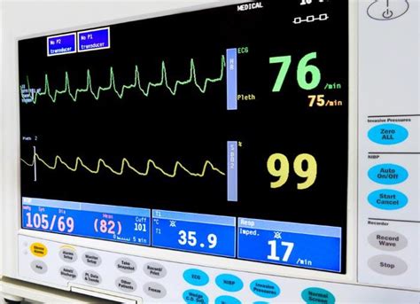
heart test machine|48 hour heart holter monitor : bespoke A Holter monitor is a machine that continuously records the heart's rhythms. The monitor is worn for 24 to 48 hours during normal activity. How the Test is Performed. . WEBAcesse sua conta no Super Portal da Hapvida Saúde, uma rede de planos de saúde com cobertura completa e preços acessíveis. Simule seu plano e cuide da saúde de quem .
{plog:ftitle_list}
Resultado da 23 de ago. de 2021 · De forma que, além da inauguração, já se faz necessário uma agenda de reuniões para abarcar com segurança todas as pessoas, carinhosamente chamadas .
A Holter monitor is a battery-operated portable electrocardiogram (ECG or EKG) device that measures and records your heart’s activity for 24 to 48 hours or longer depending . Twenty-four hour Holter monitoring is a continuous test to record your heart’s rate and rhythm for 24 hours. You wear the Holter monitor for 12 to 48 hours as you go about .A Holter monitor is a type of portable electrocardiogram (ECG). It records the electrical activity of the heart over 24 hours or longer while you are away from your healthcare provider's office. A . A cardiac computed tomography (CT) scan, also called a "CAT scan,” is a painless, non-invasive imaging test that uses X-rays to take many detailed pictures of your heart and its blood vessels. Computers can combine .

A Holter monitor is a machine that continuously records the heart's rhythms. The monitor is worn for 24 to 48 hours during normal activity. How the Test is Performed. . A Holter monitor is a battery-operated portable electrocardiogram (ECG or EKG) device that measures and records your heart’s activity for 24 to 48 hours or longer depending . The Holter Monitor test is an ambulatory ECG test used for diagnosing transient heart arrhythmias. It is quite safe and effective. Echocardiogram is a special test that uses an ultrasound machine to look at the structure and function of the heart. Chaikom/Shutterstock The images can also help them get information about:
The American Heart Association explains the common tests for heart failure and the procedures for diagnosing heart failure. Skip to main content . . A technician will use the X-ray machine to take pictures of your . A heart problem you were born with, called a congenital heart defect. Irregular results on an exercise stress test. Chest injury. Heart valve disease that needs surgery. An angiogram typically is not done until other . Nuclear Stress Test: What is it? A nuclear stress test is a minimally invasive diagnostic imaging procedure designed to evaluate the perfusion of blood through the coronary arteries to the heart muscle. Images of the heart are acquired when the heart is in a baseline resting state and again after the heart has been exercised, or stressed.Images are taken . A PET scan of the heart is a noninvasive nuclear imaging test. It uses radioactive tracers (called radionuclides) to produce pictures of your heart. Health care professionals use cardiac PET scans to diagnose coronary artery disease (CAD) and damage due to a heart attack. PET scans can show healthy and damaged heart muscle.
what machine checks your heart
Heart muscle damage. The test can show how the heart wall helps the heart pump blood. Areas of heart wall that move weakly may be damaged. Such damage might be due to a lack of oxygen or a heart attack. Heart valve disorders. An echocardiogram can show how the heart valves open and close. The test is often used to check for leaky heart valves.
Myocardial perfusion imaging (MPI) is a non-invasive imaging test that shows how well blood flows through your heart muscle. It can show areas of the heart muscle that aren’t getting enough blood flow. It can also show how well the heart muscle is pumping. This test is often called a nuclear stress test. Lack of blood flow in parts of the heart. Areas of the heart that don't show the radioactive tracer have damage from a heart attack. If you don't have enough blood flow through your heart, you may need a test called coronary angiography. This test helps health care providers see blockages in the heart arteries.
Finding out how long a wave takes to travel from one part of the heart to the next shows if the electrical activity is normal or slow, fast or irregular. Second, by measuring the amount of electrical activity passing through the heart muscle, a cardiologist may be able to find out if parts of the heart are damaged, too large or are overworked. An echocardiogram is a test that uses ultrasound to show how well your heart is working. Learn more about the echocardiogram: what it is, what it tests, types of echocardiograms, how to prepare . Cardiac troponin test: Doctors use a cardiac troponin test to help diagnose a heart attack. Troponin is a protein in the heart. If damage occurs to the heart, it sends troponin into the . Here are two easy ways to measure your heart health at home without equipment. Plus, learn the most common signs and symptoms of heart problems to look out for. . 58% of patients who took longer .
machine used to monitor heart beat
Learn about heart tests, like cardiac stress tests, echocardiograms, and electrocardiograms, and get practical tips to help you prepare. . The electrodes record the electrical activity of your heart, and the ECG machine .
A Holter monitor is a machine that continuously records the heart's rhythms. The monitor is worn for 24 to 48 hours during normal activity. . You do not need to prepare for the test. Your provider will start the monitor. You'll be told how to replace the electrodes if they fall off or get loose. Contact your provider if the monitor turns off . A cardiac MRI can help your health care team diagnose many different heart conditions, such as: Tissue damage from a heart attack. Reduced blood flow in the heart muscle to help determine whether heart artery .
how hard is fbi phase 1 test
Both evaluate how your heart performs under stress. But a nuclear stress test is a more advanced method that provides more detailed information. An exercise stress test uses only an EKG to monitor your heart . Blood tests. Certain heart proteins slowly leak into the blood after heart damage from a heart attack. Blood tests can be done to check for these proteins. . In a cardiac CT scan, you lie on a table inside a doughnut-shaped machine. An X-ray tube inside the machine rotates around your body and collects images of your heart and chest. Heart .
This heart CT test can show calcium deposits in the blood vessels. Know how the findings relate to your heart disease risk. . You lie on your back on a movable table. The table slides into a large machine shaped like a tube. Your head is outside the scanner the whole time. The exam room will likely be cool. You may be given medicine to slow . Sound waves create images of the heart in motion. This test shows the structure of the heart and heart valves and blood flow through the heart. An echocardiogram can help determine whether a ventricular assist device is the right treatment option. . If so, you'll be connected to a heart-lung bypass machine. The machine keeps oxygen-rich blood . A nuclear stress test is one of the most common tests cardiologists use to diagnose and monitor many heart conditions. The test measures blood flow to the heart at rest and after activity, or stress. . a donut-shaped imaging machine, to take pictures of your heart after the injection of a radioactive tracer. The patient is scanned at rest and .
A regular stress test uses an ECG to record your heart rate and rhythm, while a nuclear stress test captures images of your heart. The nuclear stress test uses a radioactive tracer to make a picture. The tracer is a very small amount of radioactive material that is delivered into your body through an intravenous (IV) line.Some nuclear medicine equipment has CT capabilities that help improve the images and increase the ability to combine functional imaging (nuclear medicine) and anatomic imaging (CT). . it is possible that you could experience chest pain during exercise or when a drug is given for the stress test. However, your heart will be monitored and if . Transesophageal echocardiography (TEE) is a test that produces pictures of your heart. TEE uses high-frequency sound waves (ultrasound) to make detailed pictures of your heart and the arteries that lead to and from it. . The electrodes are attached by wires to a machine that will record your electrocardiogram (ECG) to track your heartbeat .
48 hour holter monitor results
Tests are used to diagnose a heart condition or to see how healthy your heart is. Find out what to expect from some of the most common tests. . Magnetic Resonance Imaging (MRI) is a way of creating detailed images of your internal organs. Cardiac MRI uses an MRI machine to look specifically at your heart and surrounding blood vessels.Imagine a future where you can detect and diagnose heart conditions faster and with greater accuracy. With our treadmill stress test equipment, that future is here. We're delivering the accurate, intelligent insights you need to help improve patient outcomes. Get ready to experience more user-friendly and patient-friendly cardiac stress test .
On these pages you’ll find an easy-to-understand overview of seven key heart tests that your doctor may recommend. Use this information as a conversation-starter with your doctor, and as a tool in managing your own good heart health. . equipment, the test reveals whether the heart is getting enough blood flow and also determines exercise . These tests use radioactive tracers to create images of the heart. There are three types: PET Scan: It helps diagnose coronary artery disease (CAD) and heart damage after an attack. MUGA Scan: Measures ejection fraction, which shows how well your heart pumps blood. SPECT Scan: Shows blood flow and heart function at rest and during exercise, helpful for .
48 hour heart holter monitor
30 day heart monitor results
24 hour heart monitor palpitations

Resultado da 2 de fev. de 2024 · Entrar para comentar. 330 curtidas,Vídeo do TikTok de KevinMariele Conta Secundária (@kevinmariele.oficiall): "Ja doeu o .
heart test machine|48 hour heart holter monitor