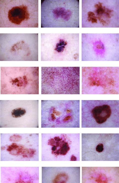
in vivo thickness measurement of basal cell carcinoma|In vivo thickness measurement of basal cell carcinoma and actinic : China In vivo thickness measurement of basal cell carcinoma and actinic keratosis with optical coherence tomography and 20-MHz ultrasound Julie Forman 2009, British Journal of Dermatology WEBThe Battle of Monte Cassino took place from 17 January 1944 to 18 May 1944. It was a series of four offensives carried out by Allied troops in central Italy (who was a key ally of .
{plog:ftitle_list}
Resultado da 11 de out. de 2020 · Olá pessoal neste vídeo mostro como fazer um corte de cabelo reto, espero que gostem não esqueçam de curtir bjssscontato .
Imaging with optical coherence tomography (OCT) has the potential to diagnose and measure depth of NMSC. Objectives: To compare accuracy of mean tumour thickness measurement in NMSC tumours < 2 mm of depth using OCT and 20-MHz high-frequency ultrasound (HFUS). The primary aims of this study were to compare accuracy of mean tumour thickness measurements in AK and BCC lesions < 2 mm of depth using 20‐MHz HFUS and .
OCT is an emergent technology that has the potential to diagnose basal cell carcinoma (BCC) in vivo and Mohs micrographic surgery offers high cure rates of .Objective: The objective was to correlate measurements of the depth of basal cell carcinomas obtained by optical coherence tomography and standard histopathologic examinations. Methods: Twenty previously scanned optical coherence tomography images of histopathologically confirmed basal cell carcinoma were reviewed. A computer-generated depth .In vivo thickness measurement of basal cell carcinoma and actinic keratosis with optical coherence tomography and 20-MHz ultrasound Julie Forman 2009, British Journal of Dermatology The definition of lateral margins in vivo needs further studies; however, ex vivo margin assessment seems convincing. There is a diagnostic role for HFUS in identifying higher recurrence risk BCC subtypes, which can help in risk stratification. . In vivo thickness measurement of basal cell carcinoma and actinic keratosis with optical .
A search of Central, Medline, Embase, CINHAL, and of Science was performed using key/MESH terms "ultrasonography" and "basal cell carcinoma" (January 2005-December 2020). We included primary studies reporting biopsy-confirmed BCCs for which the target intervention was ultrasound assessment at 15 MHz or higher frequency.
cSCC accounts for 20% of keratinocyte carcinoma (KC), with ratios of basal cell carcinoma (BCC) to cSCC ranging from 2 to 4:1. The European data on metastatic risk reveal a 2.1% cumulative incidence after a median follow-up of 15.2 months, with higher risk in males, older individuals, and certain anatomical locations . Optical coherence tomography, compared with routine histopathologic techniques, shows promise as a method for estimating the superficial thickness of basal cell carcinoma. BACKGROUND Optical coherence tomography uses advanced photonics and fiber optics to obtain high-resolution cross-sectional images and tissue characterization in real time. .
Optical coherence tomography for the characterization of basal cell carcinoma in vivo: a pilot study. . shows promise as a method for estimating the superficial thickness of basal cell carcinoma. Expand. 71. Save. . In vivo thickness measurement of basal cell carcinoma and actinic keratosis with optical coherence tomography and 20‐MHz .
Special attention has been paid to superficial basal cell carcinoma (BCC), and a number of smaller observational studies have been pub-lished. . Jemec GB (2009) In vivo thickness measurement of .
The goal of treatment for basal cell carcinoma is to remove the cancer completely. Which treatment is best for you depends on the type, location and size of your cancer, as well as your preferences and ability to do follow-up visits. Treatment selection can also depend on whether this is a first-time or a recurring basal cell carcinoma.Line-field optical coherence tomography: in vivo diagnosis of basal cell carcinoma subtypes compared with histopathology Clin Exp Dermatol. 2021 Dec;46(8) :1471-1481. . Background: Basal cell carcinoma (BCC) is the most common skin cancer in the general population. Treatments vary from Mohs surgery to topical therapy, depending on the subtype
In vivo thickness measurement of basal cell carcinoma and actinic keratosis with optical coherence tomography and 20-MHz ultrasound RCM can not only identify tumor borders by large scanning filed in superficial and early nodular basal cell carcinoma thereby determining surgery border but also perform subsequent follow‐up visits after treatment. 25 Therefore, . In vivo epidermal thickness measurement: ultrasound vs. confocal imaging. Skin Res Technol. 2004; 10 (2):136‐140.
Objectives To study the morphological features of Basal Cell Carcinoma (BCC) and measure BCC thickness by means of UHFUS examination. . OCT, optical coherence tomography; BCC, basal cell carcinoma. ** Includes ex vivo assessment. . thickness measurement of basal cell carcino- mor thickness. J Am Acad Dermatol. 1996; 32 Tormo A, Celada F .Nori S, Rius-Díaz F, Cuevas J, et al. Sensitivity and specificity of reflectance-mode confocal microscopy for in vivo diagnosis of basal cell carcinoma: a multicenter study. Journal of the American Academy of Dermatology. 2004;51(6):923–930. doi: 10.1016/j.jaad.2004.06.028.
Morphological Characteristics and Thickness of Cutaneous Basal Cell Carcinoma: A Systematic Review Alexandra aLaverde-Saad Alexe aSimard b David Nassim b Abdulhadi Jfri . HFUS provides accurate depth measurements, especially for BCCs >1 mm. The definition of lateral margins in vivo needs further studies; however, ex vivo margin as- Basal cell carcinoma is rarely lethal but can generate a high degree of disfigurement. . In vivo thickness measurement of basal cell carcinoma and actinic keratosis with optical coherence tomography and 20‐MHz ultrasound . This data indicates that in vivo reflectance confocal microscopy is a noninvasive imaging technique that has proved .The limited tissue sampling of a biopsy can lead to an incomplete assessment of basal cell carcinoma (BCC) subtypes and depth. Reflectance confocal microscopy (RCM) combined with optical coherence tomography (OCT) imaging may enable real-time, noninvasive, comprehensive three-dimensional sampling in vivo, which may improve the diagnostic accuracy and margin .
1. Introduction. Basal cell carcinoma (BCC) is the most common form of non-melanoma skin cancer. It is characterized by a slowly growing tumor originating from basal cells in the deepest layer of the epidermis, and is most common on the head, neck, and face [1].The incidence of BCC is increasing worldwide [2], leading to a considerable economic burden on .
Wallace’s group imaged multiple samples of basal cell carcinoma (BCC) in vivo . The difference between cancerous tissue and normal tissue, and the extent of the cancer invasion below the surface, are clearly visible in the THz images (Figure 2H). In a recent study, Wu and colleagues performed THz in vivo imaging in the brains of live mice . The depth of invasion by basal cell carcinoma (BCC) subtypes varies.To investigate BCC invasion depth variation by subtype and anatomic site.A prospective consecutive case series of excised BCC from 2009 to 2014 in a single Australian clinic.Descending .
In vivo thickness measurement of basal cell carcinoma and actinic keratosis with optical coherence tomography and 20-MHz ultrasound. Br J Dermatol. 2009; 160 (5):1026–1033.In vivo differentiation of common basal cell carcinoma subtypes by microvascular and structural imaging using dynamic optical coherence tomography. Exp Dermatol. 2018;27(2):156–65. 5. Ahluwalia J, Avram MM, Ortiz AE. Outcomes of Long-Pulsed 1064 nm Nd:YAG Laser Treatment of Basal Cell Carcinoma: A Retrospective Review.
Basal cell carcinoma, squamous cell carcinoma, and Merkel cell carcinoma are the three main types of nonmelanoma skin cancers and their rates of occurrence and mortality have been steadily rising . In vivo thickness measurement of basal cell carcinoma and actinic keratosis with optical coherence tomography and 20-MHz ultrasound Br. J. Dermatol. , 160 ( 5 ) ( 2009 ) , pp. 1026 - 1033 Crossref View in Scopus Google Scholar Optical coherence tomography (OCT) is a noninvasive imaging tool used in vivo in real time for diagnosis, treatment delineation and monitoring of basal cell carcinoma (BCC). Features of BCC on OCT have been widely described and reviewed. However, the diagnostic accuracy of OCT in these various applications is unclear.
Basal cell carcinoma tumor nests have been previously imaged with various optical imaging techniques. 3-7,9,13-18 The morphologic structures inside the tumor nests were clearly observed only in 2 reported studies 13,18 on ex vivo imaging of BCC in human skin using FLIM. Optical coherence tomography (OCT) is a noninvasive imaging tool used in vivo in real time for diagnosis, treatment delineation and monitoring of basal cell carcinoma (BCC). Features of BCC on OCT have been widely described and reviewed. However, the diagnostic accuracy of OCT in these various applications is unclear.

In vivo thickness measurement of basal cell carcinoma and actinic
WEB12 de dez. de 2022 · Best Close Quarters Loadout. Weapon 1. Fennec SMG. Weapon 2. Signal Sniper Rifle. One of the most difficult aspects of Warzone is clearing buildings and other close-quarter areas. The Fennec .
in vivo thickness measurement of basal cell carcinoma|In vivo thickness measurement of basal cell carcinoma and actinic