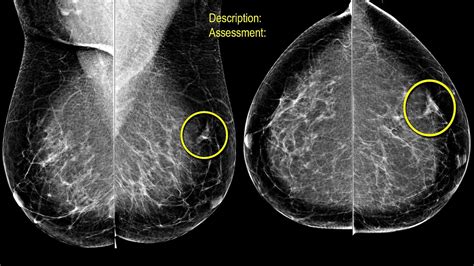
spot compression test on breast|spot compression mammogram and ultrasound : maker Magnification views are often used to evaluate micro-calcifications, tiny specks of calcium in the breast that may indicate a small cancer. Spot compression is also known as . 4 de out. de 2023 · Welcome back, Assassin’s Creed. Assassin’s Creed Mirage will be released on Oct. 5 on PlayStation 5, Windows PC, and Xbox Series X. The game was .
{plog:ftitle_list}
Murdoch Mysteries is a Canadian mystery drama television series that began in 2008. The series is based on the Detective Murdoch novels by Maureen Jennings and is set in Toronto around the turn of the 20th century.
Learn about what your mammogram results mean, including the BI-RADS system that doctors use to describe the findings they see. See moreDoctors use a standard system to describe mammogram findings and results. This system (called the Breast Imaging Reporting and Data System or BI-RADS ) . See more
hard drive test long dst failed
Your mammogram report will also include an assessment of your breast density, which is a description of how much fibrous and glandular tissue is in your breasts, . See more Magnification views are often used to evaluate micro-calcifications, tiny specks of calcium in the breast that may indicate a small cancer. Spot compression is also known as . The spot compression is just a small, plastic compression paddle that puts a little more pressure in one area, and using that we can frequently tell that something that .
Breast self-exams are important because they allow you to get to know your breasts and their “normal” appearance. If you notice abnormal symptoms or changes to your breast geography, request additional testing.
hard drive test mac
Regular mammograms take two X-ray images of each breast: top to bottom and a side-to-side view from an angle. The pictures it produces are two-dimensional, or 2D. As a . Diagnostic mammographic views may include spot compression, magnification, rolled, extended views, and true lateral views among others in order to characterize and .Spot compression and magnification views assess focal areas of interest in the breast. These views are particularly beneficial when the following are necessary1,2: Facilitate localized . Screening mammography is the primary imaging modality for early detection of breast cancer because it is the only method of breast imaging that consistently has been found .
hard drive test utility free
FIGURE 8-6 A. The actual focal spot size is determined by the electron distribution area incident on the anode. The nominal focal spot size is specified on a reference axis bisecting the field from the cathode to the anode, .
As a result, overlapping breast tissue in these pictures can hide breast cancers or make a normal spot appear to be abnormal. Tomosynthesis uses X-rays, too, but it takes more pictures from more . To explore the diagnostic efficacy of tomosynthesis spot compression (TSC) compared with conventional spot compression (CSC) for ambiguous findings on full-field digital mammography (FFDM). In . The important questions include whether the finding is new or developing, persists on spot compression, and is suspicious based on its imaging features. If the finding is suspicious, then location becomes important. . ( arrows ), indicating that the lesion is located in the superior breast. The MLO spot compression view is not helpful, but US . Ultrasound is also a useful tool for a follow-up evaluation, as well as ‘spot compression’ X-rays. Note that all mammograms are done with some breast compression, but a spot compression test uses a special plate or cone which lets you see a clearer image of a much smaller area. Margins also become clearer using spot compression.
Breast Imaging Frequently Asked Questions Update 2021. pg. 2 Screening mammography is a radiological examination to detect unsuspected breast cancer in . Additional mammographic views might include spot compression, spot compression with magnification, tangential views, or other special views. When selecting a view, the proximity of the area .
A mammogram is an x-ray of the breast. While screening mammograms are routinely performed to detect breast cancer in women who have no apparent symptoms, diagnostic mammograms are used after suspicious results on a screening mammogram or after some signs of breast cancer alert the physician to check the tissue.. Such signs and .

🎬 Check out our FREE Breast Imaging Starter Kit to learn more about the basics of mammography - https://www.mammoguide.com/Hey friends, in this video we cov. Sources of breast pain, nipple discharge or thickening skin; Why a breast has changed in size or shape; . including spot compression or spot compression with magnification, and can provide further insight into abnormalities detected during a screening. In turn, these aspects can contribute to a more accurate diagnosis or assist with .
what is spot compression mammogram
spot magnification views for mammography
Issues regarding screening for breast cancer, the role of mammography in individuals with suspected disease, and surveillance for patients with known breast cancer are discussed separately (see "Screening for breast cancer: Strategies and recommendations" and "Clinical features, diagnosis, and staging of newly diagnosed breast cancer" and .Although passing reference to the employment of coned views with compression in mammography has been made (1), the technic has been largely ignored in the literature. Gershon-Cohen (3) recommended spot-films for more accurate delineation of suspicious areas, but Wolfe (4) reported that these added little information to that of routine views. Neither author .
A screening mammogram is a low-dose imaging test that helps doctors spot changes in breast tissue. During the 10-minute procedure, a technician places your breasts—one at a time—between two imaging plates. Applying pressure to the plates produces a more detailed image with a lower dose of radiation.
spot compression vs diagnostic mammogram
It's common to be called back after a mammogram; it doesn't mean you have breast cancer. Learn why you might be called back and what other tests might be done. . The area is probably nothing to worry about, but you should have your next imaging test (mammogram and/or ultrasound) sooner than normal – usually in about 6 months – to watch .Diagnostic digital breast tomosynthesis, unilateral or bilateral (List separately in addition to 77065 and 77066) Global (Office/Freestanding) 1.59 .48 Professional (Facility) 0.86 .01 Technical (Facility) 0.73 .47 Breast Imaging: Mammography Global, Professional and Technical Payment 2021 BREAST HEALTH SOLUTIONS coding & reimbursement .Diagnostic mammography requires direct supervision.1 A diagnostic mammogram may include MLO, CC, and/or additional views to evaluate an area of clinical or radiographic concern. Additional mammographic views might include spot compression, spot compression with magnification, tangential views, or other special views.
Spot compression or a “spot view” is a mammographic technique utilized to try and spread out the breast parenchyma in an effort to decrease overlap. For a spot compression view, the technologist uses a smaller paddle which .
Compression: Ensures that the x-ray imaging system can provide adequate compression in the manual and automatic powered mode: Semiannually: Repeat Analysis: Determines the number and causes of repeated patient exposures and identifies ways to improve efficiency, reduce patient breast dose, and cut costs: Semiannually: Screen-Film Contact (N/A . Scattered fibroglanduar densities. Subcentimeter oval density lower inner quadrant left breast, middle third, which was not identified previously. ML and spot compression views as well as left breast ultrasound recommended. No suspisiously clustered microcalfications, areas of architectural distortion or axillary adenopathy.
That's because compression is part of the exam. Dr. Susan Kennedy, breast imaging radiologist, explains why compressionn is so important. MAKE AN APPOINTMENT. PAY BILL. PATIENT PORTAL. . If the breast is not well compressed, overlapping tissue can look like a mass or abnormality. This can increase the likelihood of a patient getting called . Compression helps to spread out the normal fibroglandular (dense) tissue of the breast making it easier for radiologists to see through the breast tissue and detect abnormalities that might be hidden by the overlying tissue. If the breast is not well compressed, overlapping tissue can look like a mass or abnormality.What is breast tomosynthesis? Breast tomosynthesis, also called three-dimensional (3-D) mammography and digital breast tomosynthesis (DBT), is an advanced form of breast imaging, or mammography, that uses a low-dose x-ray system and computer reconstructions to create three-dimensional images of the breasts. Breast tomosynthesis aids in the early detection .

All mammograms involve compression of the breast. Spot views apply the compression to a smaller area of tissue using a small compression plate or cone. By applying compression to only a specific area of the breast, the effective pressure is increased on that spot. This results in better tissue separation and allows better visualization of the .
February 16, 2022—According to an article in ARRS’ American Journal of Roentgenology (AJR), digital breast tomosynthesis (DBT) spot compression view could help characterize equivocal DBT findings, thus reducing further workup for benign findings. Noting that DBT spot compression increased both intrareader and interreader agreement and improved diagnostic . Digital mammography is the standard of care for screening and diagnostic imaging. When digital mammography yields equivocal findings (e.g., possible masses, subtle architectural distortions, or asymmetries), a supplementary spot compression image is obtained to reduce the superimposition of overlapping breast glandular tissue [1, 2].Digital breast tomosynthesis . The radiologist is documenting either "spot magnification radiography" or "spot compression view". the provider submitted G0206 (dx mammo producing direct digital image, unilat, all views). I am having a difficult time clarifying that the above provider verbiage supports the HCPCS code submitted.
Breast tomosynthesis (3D mammography) is an advanced mammography procedure that’s excellent at detecting cancer in dense breasts. . breast tissue. About half of women and people assigned female at birth (AFAB) have dense breasts. The denser your breast tissue, the harder it is to spot cancer on a standard 2D mammogram. Like standard . The patient was asked to return for additional mammographic views and an ultrasound. CC and MLO spot-compression views demonstrated no definite abnormality in this area (Figure 3), but a targeted ultrasound revealed a 5.5-mm spiculated mass at the 3 o’clock position (Figure 4). Diagnosis. Stage T1c, N0, M0 stage 1 left breast cancer. Discussion
hard drive test windows 8
hard drive tester hardware
Gry hazardowe darmowe to bowiem nasza pasja, którą chcemy dzielić się z innymi. Dlatego też przygotowaliśmy obszerną bazę gier kasynowych za free. Udostępniamy zarówno najpopularniejsze gry hazardowe za darmo automaty, jak i te znacznie .
spot compression test on breast|spot compression mammogram and ultrasound