water drop test veress needle|veres needle pressure : factory The various Veress needle safety tests are not a sensitive indicator of the proper placement of the Veress needle. The Veress intraperitoneal pressure (i.e., <10 mmHg) is a . WEBA atriz pornô Elisa Sanches atualizou as redes sociais nesta última sexta-feira (5) e publicou um vídeo picante e ousou com a cena que contou até com plateia.
{plog:ftitle_list}
WEBIf you want to experience games that are at the bleeding edge of mobile gaming .
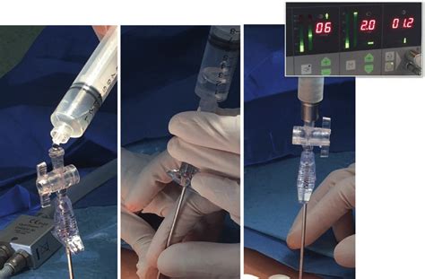
Hanging drop test – involves placing a drop of water on the open end of the Veress needle and the abdominal wall is elevated. If the needle is correctly positioned, the water should disappear down the shaft.
An unobstructed free intraperitoneal position for the Veress needle is verified by easy irrigation of clear saline in and out of the peritoneal space and by the hanging drop method where the .
veress needle technique
veress needle surgery
Abstract. Study objective: To determine the reliability of four commonly used tests to confirm the placement of the Veres needle during closed laparoscopy and their ability to determine other . The various Veress needle safety tests are not a sensitive indicator of the proper placement of the Veress needle. The Veress intraperitoneal pressure (i.e., <10 mmHg) is a . The laparoscopic entry techniques and technologies reviewed in formulating this guideline included the closed (Veress . We present a meta-analysis comparing Veress needle entry with direct trocar entry in gynecologic surgery. Our results demonstrate that the use of Veress needle entry is .
In women requiring intra-abdominal surgery in pregnancy, Veress needle insufation at the umbilical site can be. fl. employed until 14 weeks gestation (if there are no contraindi-cations), . Entry using the optical Veress needle is also called “minilaparoscopy.” A modified Veress needle of 2.1 mm diameter and a 10.5-cm-long cannula are used, allowing . A hanging drop test was performed by elevating the retractors to observe a drop of fluid placed on the hub of the Veress needle getting sucked in Confirmatory test: The Veress . The following tests that attempt to determine the correct intra-abdominal placement of the Veress needle have been described: the double click sound of the Veress needle; the Palmer’s test (aspiration test); the hanging drop of saline test ; the “hiss” sound test ; the syringe test [35–38]; and the pressure profile test, of which the .
4. Previously recommended Veress needle safety checks or tests, such as the saline drop test and aspiration for fluid, have not been found to confirm position and therefore are no longer recom-mended as best practice (I-A). 5. Wiggling the Veress needle from side to side should be avoided; this can increase the risk of complications (II-1E). 6. Tests and techniques for determining intraperitoneal placement of the Veress needle include the double-click sound/acoustic test of the Veress needle as it traverses the fascia and the peritoneum, the aspiration test, the . In our assessment, incorrect placement of the needle seemed to be the major cause of these injuries.26 In many discussions regarding the tests to determine the correct needle insertion, there seemed to be agreement that the ‘drop’ test (intra-abdominal pressure vs saline within the Veress needle) seemed to be the most reliable.
In our assessment, incorrect placement of the needle seemed to be the major cause of these injuries. 26 In many discussions regarding the tests to determine the correct needle insertion, there seemed to be agreement that the ‘drop’ test (intra-abdominal pressure vs saline within the Veress needle) seemed to be the most reliable. Results: The average time from cleaning umbilicus again to Veress needle tip in peritoneum was 1 min 40 s (25 s–7 min). Out of the 4228 patients in whom no adhesions were observed at first trocar entry (Group 1), the Veress needle insertion was successful at first attempt in 3829 (90.5%) patients, at second attempt in 322 (7.6%) and at third attempt in 30 . Hanging drop test: Place a few drops of fluid (saline or sterile water) over the Veress needle allowing a drop to hang at the head. Then slightly lift the abdominal wall. If the Veress needle is correctly located inside the peritoneal cavity, the hanging drop will be sucked inside because of the negative pressure of the peritoneal cavity.
Customer Service: 1-800-448-6506 International Sales: 1-315-797-8375 Fax: 1-800-438-3051 CONMED.com [email protected] CONMED Corporation 525 French Road, Utica, NY 13502The needle was introduced as a safer technique to give patients such pneumothoraces. It was not until 1938, when he published his invention in the German literature, that the needle became more broadly known outside of Hungary. [4] Raoul Palmer introduced the use of the Veress needle in laparoscopy to establish a pneumoperitoneum. [5]needle, the only reliable indicator is Veress Intraperitoneal Pressure (VIP) below ≤ 8 mmHg. Other Veress placement tests (e.g. double-click or saline hanging-drop) are not required while swinging the needle is hazardous and must be avoided (Grade II-1/A). To ensure adequate pneumoperitoneum, transient highThis video demonstrates laparoscopic veress needle. Dr R K Mishra explained veress needle. A Veress needle is a spring-loaded needle used to create pneumoper.
veress needle positioning procedure
The Veress needle was introduced through the umbilical scar by giving a supraumbilical curvilinear skin incision. In all patients abdominal wall was lifted with nondominant hand or by the assistant to facilitate safe and easy introduction of Veress needle. The entry into the abdominal cavity was confirmed by double click sound and later on by . The safety profile of the Veress needle has always been questioned over the years. We share our experience with the exclusive use of Veress needle in consecutive 2631 patients for pediatric laparoscopy over the past 18 years. . A hanging drop test and aspiration were done to confirm intra-peritoneal placement in all cases. A maximum of two .Use of the Veress needle to obtain pneumoperitoneum prior to laparoscopy . 2.6 Test placement Once the Veress needle tip is in the peritoneal cavity, the clinical may consider the aspiration and saline drop tests. It should be noted that these tests have moderate sensitivity and specificity for correct entry only. History. After its invention in 1936, the Veres needle lay dormant for many years. The video computer chip was developed in 1986, which allowed projection of the laparoscopic image on to a screen 1.This led to the implementation of laparoscopic surgery through the 1990s, and the need for methods to establish pneumoperitoneum revived the Veres needle.
Abdominal Veress needle insertion is commonly performed to generate a pneumoperitoneum during laparoscopy. Various safety tests are conducted to confirm accurate needle tip positioning into the abdominal cavity. . The principle of the NPV is identical to the hanging drop test, falling water column test, or manometer test in terms of detecting . Previously recommended Veress needle safety checks or tests, such as the saline drop test and aspiration for fluid, have not been found to confirm position and therefore are no longer recommended as best practice (I-A). 5. Wiggling the Veress needle from side to side should be avoided; this can increase the risk of complications (II-1E). 6
The aspiration test. The Veress needle should be aspirated with a 5-mL syringe after placement. Aspiration of fresh blood fresh blood through the needle suggests that the tips had entered a vein. 2 • The drop test. A drop of saline is placed in the opened hub of the Veress needle, and the abdominal wall is lifted.
The most common access site for insufflation with a Veress needle is at the umbilicus since there is no fat or muscle between the skin and the peritoneum; however, entry at Palmer’s point (see below) is a much safer location. . If you are not comfortable with the insufflation pressures or the results of the saline drop test, do not continue . Standard saline drop test was employed to test for correct placement of the Veress needle. Clinical observations regarding ease of insertion, number of attempts made to place Veress needle in the peritoneal cavity and hemopneumothorax were made immediately after VNI, and observations concerning intra-abdominal bleed from parities and intra . Safe insertion of the Veress needle during laparoscopy relies on the surgeons’ technical skills in order to stop needle insertion just in time to prevent overshooting in the underlying organs. 1. VERESS NEEDLE AND SAFETY OF VERESS NEEDLE INSERTION The Veress needle, a pivotal instrument in laparoscopic surgery, serves the crucial role of initiating pneumoperitoneum by safely introducing CO2 into the abdominal cavity. Originating in the late 1940s, its adoption marked a significant advancement in surgical technique.


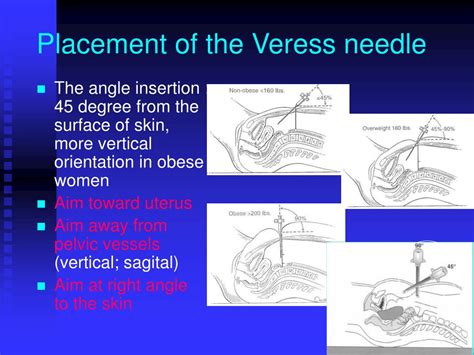
Measurements and main results: Caudal displacement of the umbilicus before insertion of the veress needle allows for a median displacement of 6 cm between the site of entry and the common iliac vessels. An entry pressure of less than 9 mm Hg is suggestive of successful intraperitoneal entry. The left upper quadrant should be used in specific .The Veress needle is always inserted at a 90-degree angle at Palmer’s point regardless of BMI. Once pneumoperitoneum has been established, the trocar can be placed blindly (as demonstrated in the video) or using an optical trocar, where the surgeon visualizes the fascia, rectus muscles, and peritoneum on entry to the abdominal cavity.If the tip of the Veress needle is anywhere else the hanging drop test will be negative. Once it is confirmed that the Veress needle is inside the abdominal cavity the tubing of the insufflator is attached and flow is started. Hanging drop test Measurement of Intra-abdominal Pressure
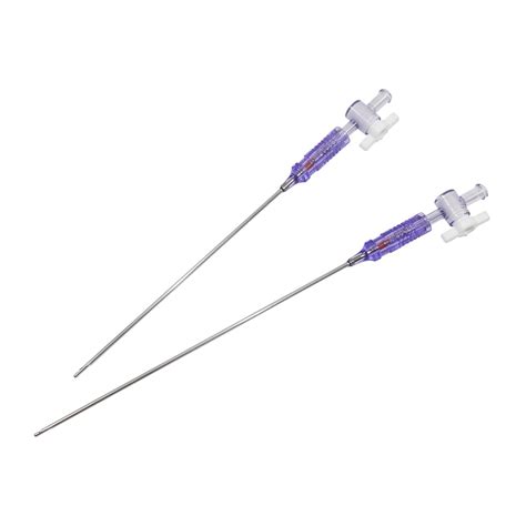
The video shows the steps of using a veress needle.SurgSci is a professional laparoscopic instruments manufacturer and supplier in China. Veress neeedles are.
veress needle positioning diagram
A double click and initial low pressure ≤9mmHg on CO 2 insufflation were taken as indicators of intra peritoneal position of veress needle. 8 No other test was used in routine. View Show abstract

veress needle
veres needle pressure
veres needle gas pressure
webZACA GRAVA VÍDEO AGRADECENDO AOS INTERNAUTAS O PRIMEIRO LUGAR EM AUDIÊNCIA DO 'PORTAL DO ZACARIAS' NO AMAZONAS E NA REGIÃO NORTE. 'PORTAL DO ZACARIAS' TAMBÉM JÁ É O DÉCIMO PORTAL MAIS ACESSADO DO BRASIL. OS DADOS SÃO DO IVC . Porcos não transpiram, então de onde veio a .
water drop test veress needle|veres needle pressure