what medical test shows soft tissue in the back|benign soft tissue cancer imaging : distributing PET-CT detects positron emission decay from an administered radioisotope and generates an image of the entire body. The amount of radiotracer activity within the cells reflects metabolic . 6 dias atrás · Por GIGA-SENA. O sorteio do concurso 6373 ocorreu no dia 23 de fevereiro de 2024 e o prêmio principal foi estimado em R$ R$ 1.300.000,00 (um milhão e trezentos mil reais) para quem acertar o resultado da Quina 6373. Quem acertar a QUADRA com 4 (quatro) números, o TERNO com 3 (três) números ou o DUQUE com 2 (dois) números .
{plog:ftitle_list}
Melissa Devassa (@melissa.devassa) no TikTok |393K curtidas.49.9K seguidores.54 anos, 1,83 de altura, libriana 5 filhos, 2 netas Cinquentando com alegria.Assista ao último vídeo de Melissa Devassa (@melissa.devassa). Passar para o feed de conteúdo. TikTok. Carregar . Entrar.
An X-ray, also called a radiograph, sends radiation through the body. Areas with high levels of calcium (bones and teeth) block the radiation, causing them to appear white on the image. Soft tissues allow the radiation to pass through. They appear gray or black on the image. An X-ray is the fastest and most accessible . See more
An MRI, or magnetic resonance imaging, uses a powerful magnet to pass radio waves through the body. Protons in the body react to the energy and create highly detailed pictures of the body’s structures, including soft tissues, nerves and blood vessels. Unlike X . See moreA CT scan, or computed tomography scan, sends radiation through the body. However, unlike a simple X-ray study, it offers a much higher level of detail, creating computerized, 360-degree views of the body’s structures. CT scans are fast and detailed. They . See moreA CT scan may be recommended if a patient can’t have an MRI. People with metal implants, pacemakers or other implanted devices shouldn’t have an MRI due to the powerful . See morePET-CT detects positron emission decay from an administered radioisotope and generates an image of the entire body. The amount of radiotracer activity within the cells reflects metabolic .
MRI is the most accurate modality for diagnosing soft tissue masses because it can provide further information regarding adjacent anatomic structures, presence of necrosis, border definition,.
mri for benign soft tissue
benign soft tissue pain
Medical pathologists examine tissue samples under a microscope to determine if the tumor is benign or cancerous. Tests show I have a benign soft tissue tumor. Should I be . A spine X-ray is an imaging test that uses electromagnetic waves to take detailed pictures of the bones in your neck and back. You might need spinal X-rays if you were born .This test is often done if the doctor suspects a soft tissue sarcoma in the chest, abdomen (belly), or the retroperitoneum (the back of the abdomen). This test is also used to see if the . Space-occupying soft-tissue lesions are frequently detected on MRI studies, either as incidental findings or during the evaluation of palpable abnormalities or focal symptoms. In this setting, the exclusion of malignancy is .
A sarcoma is a type of cancer that develops in bone or soft tissues like muscle, nerves, fat, fibrous tissues, tendons, or blood vessels. Sarcomas can grow anywhere in the body, but they most often appear as a lump or bump on the . A CT scan of the back may view one or more of the three areas of the spine: the cervical spine (neck), thoracic spine (middle back), and lumbar spine (lower back). Doctors .Less dense soft tissues and breaks in bone let radiation pass through, making these parts look darker on the X-ray film. You will probably be X-rayed from several angles. If you have a fracture in one limb, your doctor may want a .

Soft Tissue Masses: Diagnosis and Surgery for Benign and Cancerous Tumors . Diagnostic tests. Patients presenting with soft tissue masses are evaluated and their clinical history taken. Diagnostic tests might include X-ray, magnetic . The structures that are imaged in soft-tissue bedside ultrasound are primarily the skin, subcutaneous tissue, fascia, and muscle. The skin consists of two layers: the superficial epidermis and the deeper, thicker dermis. The .
benign soft tissue diagnosis
A doctor may use an ultrasound scan as one of the first tests to diagnose and assess a soft tissue sarcoma. An ultrasound can help doctors determine the nature of the lump and plan the most .Tests for soft tissue sarcoma. You usually have a number of tests to check for soft tissue sarcoma. Soft tissue sarcomas are cancers that start in the connective and supporting tissues of the body. These include the: fat. muscle. blood vessels. deep skin tissues. nerves. tendons and ligaments. tissues around the joints. The bones are also a .
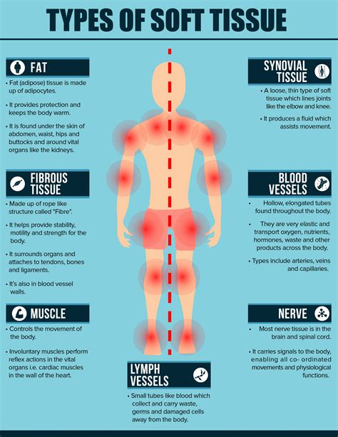
Ultrasound is a noninvasive imaging test that shows structures inside your body using high-intensity sound waves. An ultrasound picture is called a sonogram. . Sound waves bounce off structures inside your body and back to the probe, which converts the waves into electrical signals. . Soft-tissue masses. Organs (liver, kidney or prostate).It depends on the amount of X-rays that penetrate the tissues. The soft tissues in the body (like blood, skin, fat, and muscle) allow most of the X-ray to pass through and appear dark gray on the film. A bone or a tumor, which is denser than soft tissue, allows few of the X-rays to pass through and appears white on the X-ray.A sarcoma is a type of cancer that starts in tissues like bone or muscle. Bone and soft tissue sarcomas are the main types of sarcoma. Soft tissue sarcomas can develop in soft tissues like fat, muscle, nerves, fibrous tissues, blood vessels, or deep skin tissues. They can be found in any part of the body. Soft tissue sarcoma is a rare type of cancer that starts as a growth of cells in the body's soft tissues. The soft tissues connect, support and surround other body structures.
Computerized Film Thickness Tester Brand manufacturer
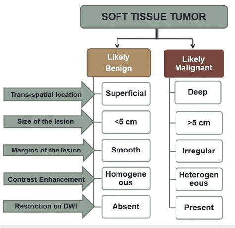
Clinical History: The clinical history section provides a brief description of the patient’s medical history relevant to the tissue sample that the pathologist is examining. Clinical Diagnosis (Pre-Operative Diagnosis): The clinical diagnosis describes what the doctors are expecting before the pathologic diagnosis. Imaging tests. A whiplash injury doesn't show on imaging tests. But imaging tests can rule out other conditions that could be making your neck pain worse. Imaging tests include: X-rays. X-rays of the neck taken from many angles can show broken bones, arthritis and other issues. CT scan. OBJECTIVE. A wide spectrum of space-occupying soft-tissue lesions may be discovered on MRI studies, either as incidental findings or as palpable or symptomatic masses. Characterization of a lesion as benign or indeterminate is the most important step toward optimal treatment and avoidance of unnecessary biopsy or surgical intervention. CONCLUSION. The . Introduction. Ultrasonography is an excellent imaging method in the evaluation of a palpable superficial soft-tissue mass. The advantages of US include high-spatial-resolution capabilities, portability, easy access, low cost, comparison with the contralateral side, Doppler US, and, importantly, the ability to combine physical examination findings and patient history during .
A soft tissue sarcoma is a cancerous tumor that can grow on soft connective tissues in your body, including muscles, nerves, blood vessels, and the lining of your joints. A computerized tomography scan, also called a CT scan, is a type of imaging that uses X-ray techniques to create detailed images of the body. It then uses a computer to create cross-sectional images, also called slices, of the bones, blood vessels and soft tissues inside the body. CT scan images show more detail than plain X-rays do.The American Cancer Society estimates about 13,190 new soft tissue sarcomas will be diagnosed in the United States this year. For comparison, more than 265,000 cases of breast cancer and nearly 270,000 cases of prostate cancer .
In medical reports, “soft tissues are unremarkable” serves as a valuable piece of information for doctors in their diagnostic process. It aids in forming accurate diagnoses by eliminating concerns related to soft tissue abnormalities, narrowing down potential health issues, and directing further examinations if needed. Common Scenarios MRI, which uses powerful magnets to produce 3-D anatomic images, is a high-contrast resolution modality that can determine changes in the tissue quality.
Along with imaging, your medical provider might order blood tests and conduct a physical exam to pinpoint the root cause and give you the best treatment possible. Imaging Tests Used for Diagnosing Muscle Disorders. Your medical provider might order one or more imaging tests for a closer look at your muscles, joints and bones. OBJECTIVE. Soft-tissue masses derive from a wide spectrum of tissues, and it may be difficult to differentiate nonneoplastic from neoplastic as well as benign from malignant lesions, to say nothing of making a single histologic diagnosis on the basis of imaging. The purpose of this article is to discuss optimal imaging protocols and reporting of soft-tissue . An MRI scan is an imaging test that creates detailed pictures of the soft tissue around the spine. Information. . back and neck pain are not caused by a serious medical problem or injury. Low back or neck pain often gets better on its own with time. Soft tissue sarcomas affect the soft tissue, such as fat or muscle. . Imaging tests: A doctor may use imaging scans such as a CT scan, . whether the cancer has come back;

Diagnostic ultrasounds use sound waves to make pictures of the body. Ultrasound, also called sonography, shows the structures inside the body. The images can help guide diagnosis and treatment for many diseases and conditions. Most ultrasounds are done using a device outside the body. However, some involve placing a small device inside the body. Tests and procedures used to diagnose soft tissue sarcoma include imaging tests and procedures to remove a sample of cells for testing. . They might help show the size and location of the soft tissue sarcoma. Examples include: X-rays. CT scans. MRI scans. Positron emission tomography (PET) scans. . medical social worker, clergy member or . Neck masses in adults require organized diagnostic methods to determine if they are malignant or benign, with imaging and fine-needle aspiration biopsy being key tools.
benign soft tissue cancer imaging
These tests typically include MRIs or CTs. The scans can show how extensive the tumor is and if it has spread. The next step is a biopsy. Since sarcomas are rare, interpretation of the biopsy is crucial. It is important that you receive a diagnosis from a team of doctors that is highly experienced in the diagnosis and treatment of soft tissue . What an X-Ray Doesn’t Show. X-rays are great to check for broken bones or rotting teeth, but other imaging tests are better if you have something happening with the soft tissue parts of your .
Soft tissue masses or lesions are a common medical condition seen by primary care physicians . Plain radiographs are of limited use for the evaluation of soft tissue masses and usually show only soft tissue shadowing. But they can show calcifications as well as bony involvement in the form of osteolysis, cortical erosion or periosteal .
WEBAcompanhantes Macapá (AP) Está em Macapá (AP) e precisa de uma companhia para viver uma noite especial? Então, acesse a plataforma Erosguia e escolha, dentre .
what medical test shows soft tissue in the back|benign soft tissue cancer imaging