testing soft tissue of the neck|soft tissue neck : chain store Portable anterior–posterior (AP) and lateral soft-tissue neck x-rays provide basic information about the airway, as described earlier. Like a focused assessment with sonography for trauma (FAST), the soft-tissue neck film should be assessed by the . See more Herbst Kurse Online. Bodyfit morgens 8:30 – 9:30 Uhr. Bodyfit abends 18:30 – 19:30 Uhr. Die Kurse finden 10x statt und können bei der Krankenkasse zur Bezuschussung eingereicht werden. Der Preis für den Kurs liegt bei .
{plog:ftitle_list}
80% OFF. Desconto de até 80% OFF em Acessórios selecionados na Santa Lolla. Não é necessário aplicar cupom de desconto Santa Lolla em sua compra. Válido para .
Imaging of soft tissues of the neck can be essential in the evaluation of patients with a variety of chief complaints, including neck trauma, ingested or aspirated foreign body, nontraumatic neck pain and swelling, dysphagia and voice change, visible or palpable mass, and central nervous system complaints with . See moreGiven the range of potential pathology discussed earlier, it should come as little surprise that no single clinical decision rule can be used to . See more
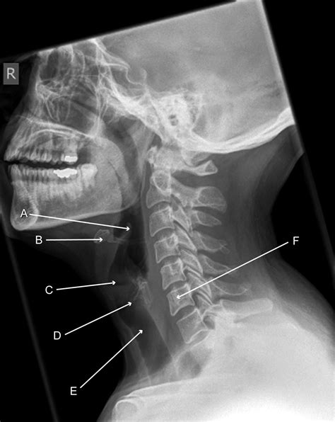
Plain x-ray (Figures 4-1 and 4-2) provides limited information about the soft tissues of the neck. X-ray relies on differentiation of adjacent structures using four basic tissue densities: air, fat, water (which includes soft tissues, both solid organs such as muscle and fluids . See morePortable anterior–posterior (AP) and lateral soft-tissue neck x-rays provide basic information about the airway, as described earlier. Like a focused assessment with sonography for trauma (FAST), the soft-tissue neck film should be assessed by the . See more
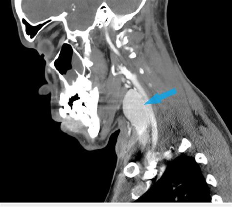
CT scans are preferred for neck imaging for several reasons: Detailed Imaging: CT scans provide detailed images of bones, soft tissues, and blood vessels in the neck area, .A CT Neck (Soft Tissue) is an exam that takes very thin slice (3.5mm) images of the neck, starting from just above the ears and ending just below the clavicles (collar bone). This allows .A CT scan of the neck can give your doctor information about your neck, throat, and tonsils and other structures in the neck area. Why is this test done? A CT scan of the neck can help find . A cervical MRI scans the soft tissues of your neck and cervical spine. The cervical spine is the portion of your spine that runs through your neck. A cervical spine MRI scan is .
The soft tissue structures of the neck that are evaluated include nasopharynx, oropharynx, laryngopharynx, thyroid, lateral pharyngeal space, and other soft tissues. The study is .
When you receive a doctor’s order for a medical test for your neck, you’ll see it written as a CT scan for the cervical spine. This is the top section of your spine that passes .
This section of the website will explain how to plan for an MRI soft tissue neck scans, protocols for MRI soft tissue neck, how to position for MRI soft tissue neck and indications for MRI soft . Soft tissues include: blood vessels. skin. fat. muscles. Read more: Vertebrae of the neck » Why is a neck X-ray performed? Your doctor may request a neck X-ray if you have a neck. MRI is a tool for detecting, characterizing, and monitoring tumors and cysts in the neck. It can help differentiate benign from malignant tumors and is particularly useful for .The soft tissue structures of the neck that are evaluated include nasopharynx, oropharynx, laryngopharynx, thyroid, lateral pharyngeal space, and other soft tissues. The study is best done with intravenous contrast. . Your patient may need a blood test (creatinine level) to check renal functions prior to restarting the medication. The CT .
This surgery is called a neck dissection and is usually (but not always) done at the same time as the primary site surgery. If the treatment plan calls for radiation therapy, the neck may be treated with radiation therapy, too. Neck dissection . A neck MRI is a medical imaging procedure that uses magnets and radio waves to create detailed images of the neck and its internal structures. Unlike X-rays and CT scans, MRIs do not use ionizing radiation, which makes them a safer option for imaging soft tissues.MRI (magnetic resonance imaging) is a test that uses a magnetic field and pulses of radio wave energy to make pictures of the organs and structures inside the body. An MRI can give your doctor information about your neck, throat, tongue, voice box (larynx), tonsils, and other structures in the neck area. Fascia is an internal connective tissue which forms bands or sheets that surround and support muscles, vessels and nerves in the body.. In the neck, these layers of fascia not only act to support internal structures, but also help to compartmentalise structures of the neck. There are two fascias in the neck – the superficial cervical fascia and the deep cervical fascia.
There is a risk of damage to cells or tissue from being exposed to radiation, including the small amounts used in CTs, X-rays, and other medical tests. Over time, exposure to radiation may cause cancer and other health problems. . Enter H055 in the search box to learn more about "CT Scan of the Neck: About This Test". Current as of: July 31 . This imaging test uses radio waves and a magnetic field to make detailed 3D images. Besides bone injuries, MRI scans can show some soft tissue injuries, such as damage to the spinal cord, disks or . They include numbness or dizziness, and rarely damage to spinal tissues. Massage. Neck massage may provide short-term relief of neck pain from . Introduction. Ultrasonography is an excellent imaging method in the evaluation of a palpable superficial soft-tissue mass. The advantages of US include high-spatial-resolution capabilities, portability, easy access, low cost, comparison with the contralateral side, Doppler US, and, importantly, the ability to combine physical examination findings and patient history during .

A CT Neck (Soft Tissue) is an exam that takes very thin slice (3.5mm) images of the neck, starting from just above the ears and ending just below the. . These are called scout images and are used to map the area for testing. During this, you will feel the table move, but you will not be touched. The scanner will, with recorded messages, ask .Soft tissue injuries in the neck. There are numerous soft tissues that attach to the neck, including muscles, tendons, and ligaments. These soft tissues all work in tandem to support your neck and head. At the same time, they also enable movement in your neck. A neck strain or sprain occurs when one or more of these soft tissues is stretched . Treatment for soft tissue injuries depends on several factors, including:. the severity; the type of injury; the particular joint, muscle, or limb affected; People can often self-treat mild soft .
A new neck mass is a relatively common head and neck problem. There often are no associated symptoms other than the recognition of a new "lump" noted incidentally on palpation while grooming or noticed by another individual. The mass may be the only manifestation of a serious and potentially malignant pathology, especially in the adult population.Study with Quizlet and memorize flashcards containing terms like Bleeding from soft-tissue injuries to the face is MOST effectively controlled with: A. direct pressure using dry, sterile dressings. B. pressure dressings and chemical ice packs. C. ice packs and elevation of the patient's head. D. digital pressure to an adjacent pulse point., Facial injuries should be .
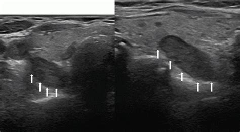
ultrasound soft tissue neck
soft tissues of the neck x ray
A neck X-ray, also known as a cervical spine X-ray, is an X-ray image taken of your cervical vertebrae. . Soft tissues include: blood vessels; . An X-ray is a common imaging test that can help .
21510 For code 21510, go to CPT index main term Incision and subterm See also Incision and Drainage. Then, go to CPT index main term Incision and Drainage, subterm Thorax, and qualifier Deep. Verify the code in the Incision subcategory of the Neck (Soft Tissues) and Thorax category in the Musculoskeletal System subsection of the Surgery section.CT-guided needle biopsy: If a lung biopsy is needed to check for cancer spread, this test can also be used to guide a biopsy needle into the mass (lump) to get a tissue sample to check for cancer. Magnetic resonance imaging (MRI) Like CT scans, MRI scans show detailed images of soft tissues in the body. But MRI scans use radio waves and strong .

soft tissues in neck ct
Excision of fascial or subfascial soft tissue tumors involves the resection of tumors confined to the tissue within or below the deep fascia but not involving the bone. These tumors are usually benign, are often intramuscular, .
soft tissue scan for neck
Workup for a neck mass may include a complete blood count; purified protein derivative test for tuberculosis; and measurement of titers for Epstein-Barr virus, cat-scratch disease, cytomegalovirus .This test is often done if the doctor suspects a soft tissue sarcoma in the chest, abdomen (belly), or the retroperitoneum (the back of the abdomen). This test is also used to see if the sarcoma has spread to the lungs, liver, or other organs.It depends on the amount of X-rays that penetrate the tissues. The soft tissues in the body (like blood, skin, fat, and muscle) allow most of the X-ray to pass through and appear dark gray on the film. A bone or a tumor, which is denser than soft tissue, allows few of the X-rays to pass through and appears white on the X-ray.MRI can provide very detailed images of soft tissues such as the thyroid gland and nearby lymph nodes. However, an ultrasound of the neck is usually the first test done to look at the thyroid. MRI might also be used to look for cancer spread to other parts of the body, although this is less common. Positron emission tomography (PET) scan
The provider performs real–time ultrasound examination of the soft tissues of the head and neck and records and saves the images for later review. For clinical responsibility, terminology, tips and additional info start codify free trial.
soft tissue neck radiology
Ultrasound enables healthcare providers to “see” details of soft tissues inside your body without making any incisions (cuts). And unlike X-rays, ultrasound doesn’t use radiation.. Although most people associate ultrasound with pregnancy, healthcare providers use ultrasound for many different situations and to look at several different parts of the inside of your body.Whiplash is an injury caused by the neck bending forcibly forward and then backward, or vice versa. . To protect your loved one, please do not visit if you are sick or have a COVID-19 positive test result. Get more resources on masking and COVID-19 . Large magnets and a computer make detailed images of organs and soft tissue structures in . A cervical MRI scans the soft tissues of your neck and cervical spine. The cervical spine is the portion of your spine that runs through your neck. A cervical spine MRI scan is used to help diagnose:
Neck pain is a common presenting symptom in primary care, with an incidence of 10.4% to 21.3% per year. 1 It is the fourth leading cause of disability worldwide. 1 The prevalence of neck pain is .The CT scan is an x-ray test that makes detailed cross-sectional images of your body. A CT scan of the head and neck can provide information about the size, shape, and position of a tumor, see if it's growing into nearby tissues, and can help find .

webFor this dino, there are 2 methods (Honest). Here they are: METHOD 1: The Trap. The usual. 2x2 stone trap (4 stone floors, 16 stone walls and 4-6 ramps), cost efficient and simple. Lead the spino into the trap and tranq it down. When taming, make sure to watch its torpor as it drains quite fast! METHOD 2: Trapless.
testing soft tissue of the neck|soft tissue neck