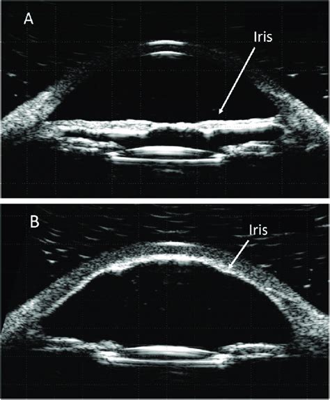
ubm scleral thickness measurement|ubm technique : wholesalers UBM can be used to measure scleral thickness, and our results support the finding that patients with uveal effusion syndrome have abnormally thick sclera. Compared with MRI, UBM may be a more accurate and precise method of measuring scleral thickness. 5 de jul. de 2021 · Na noite deste domingo,5, um vídeo foi enviado por um leitor do ' PORTAL DO ZACARIAS ' que registraram momentos chocantes. Um homem que não .
{plog:ftitle_list}
29 de mai. de 2021 · Suécia e Finlândia se enfrentam neste sábado (29) a partir das 13h (horário de Brasília), na Friends Arena, em jogo amistoso. Disputando a Eurocopa, equipes se preparam para entrar em campo .
UBM can be used to measure scleral thickness, and our results support the finding that patients with uveal effusion syndrome have abnormally thick sclera. Compared with MRI, UBM may be a more accurate and precise method of measuring scleral thickness. The in vivo methods to measure scleral thickness include ultrasound biomicroscopy (UBM) and OCT. While authors have reported scleral thickness measured using UBM, there are practical limitations in the technique as this is dependent on a technician with a high level of expertise, requires a patient to be in the supine position, and is a contact . UBM was used to measure scleral thickness in five subjects with uveal effusion syndrome and five matched controls. We also used MRI to measure scleral thickness in three subjects. Results. The mean thicknesses for eyes with uveal effusion syndrome versus control eyes were 0.65 ± 0.08 mm and 0.55 ± 0.05 mm, .The first measured ST on the projection of photographic slides of trans-sectioned eye bank eyes,14 and the second using UBM in living eyes included episcleral tissues in the measurements, which resulted in higher values for ST.15 The scleral spur is an important landmark for orientation and measuring distances in UBM images of the .
Anterior scleral thickness also showed significant variations with measurement location, and a location by meridian interaction (both p < 0.001), but the average thickness of the nasal (mean .Sclera . UBM is only able to examine the area of the sclera anterior to the equator due to the area limited by the scleral shell. . imaging and biometry of 2-D corneal cross-sections instead of at individual positions thus giving a more precise measurement of corneal thickness. 12. UBM has been used for assessment of the cornea following . Using UBM measurement techniques, Oliveira et al. found the scleral thickness at 3.0 mm post limbus to be 506.5 ± 65 µm in 140 eyes [21]. . With reference to in vivo measurement of scleral . Scleral spur identified in the area where the radiopaque shadow of the sclera converges with the radiolucent shadow of the cornea. . Studies have demonstrated the use of VHF digital UBM for measuring the flap thickness and residual stromal thickness (RST) .
To validate scleral thickness measurements by ultrasound biomicroscopy (UBM) vis a vis anterior segment optical coherence tomography (ASOCT). Methods: This case-control study analyzed 50 eyes of 50 patients with CSCR (cases) and compared it with that of 50 eyes of 50 age- and gender-matched controls.
A previous study showed that UBM may be a more accurate and precise method for the measurement of scleral thickness than magnetic resonance imaging; the study found mean thicknesses values of 0.65 . We found that the mean thickness of the sclera near the scleral process was 0.76 mm by UBM. A previous study showed that UBM may be a more accurate and precise method for the measurement of scleral thickness than magnetic resonance imaging; the study found mean thicknesses values of 0.65±0.08 and 0.55±0.05 mm for eyes with UES and control . ACD and LT measurements obtained using immersion A-scan were significantly greater than with UBM image analysis with mean differences of 0.52 mm and 0.62 mm, respectively (p < 0.001).Immersion A-scan and UBM measurements were moderately correlated (r = 0.70 and 0.64, p < 0.001). ACD and LT measurements obtained using CV overlay were . To use Optical Coherence Tomography (OCT) to measure scleral thickness (ST) and subfoveal choroid thickness (SFCT) in patients with Branch Retinal Vein Occlusion (BRVO) and to conduct a .
vehicle battery voltage drop test
However, scleral thickness measurements using MRI were less accurate and precise compared to UBM, likely as a result of movement artifacts. Surgical Treatment Methods Full-thickness and partial-thickness sclerectomy is the preferred surgical approach for most vitreoretinal specialists.
Scleral thickness was determined through three methods: direct measurements with a digital caliper, image projection, and ultrasonic biomicroscopy (UBM). Results The average scleral surface areas in canine and equine eyes were approximately .Average thickness was measured in each region, giving 60 thickness measurements/eye. Scleral geometric features were correlated with globe axial length. Results: : Group mean thickness over the whole sclera was 670±80 µm (mean±SD). Maximum thickness occurred at the posterior pole of the eye, with mean thickness of 996±181 µm. There are newer UBM devices that overcome some of these limitations, but I have limited experience with those.” . “You can get corneal thickness measurements using anterior segment OCT,” notes Dr. . Ultrasound biomicroscopy (UBM) is a good investigation for measuring the scleral thickness of the anterior segment, although it only visualizes 3.0 mm posterior from the scleral spur. Therefore, for posterior .

However, scleral thickness measurements using MRI were less accurate and precise compared to UBM, likely as a result of movement artifacts. Surgical Treatment Methods Full-thickness and partial-thickness sclerectomy is the preferred surgical approach for most vitreoretinal specialists.
AST, Norman et al ., [15] used 3-dimensional microMR I to measure in vitro scleral thickness for circumferential slices of whole eyes of normal and glaucomatous patients. The slices wereDownload scientific diagram | Original image: Measurement of AST: ASOCT (Top)-White arrows from top to bottom showing structures as labelled. UBM (Bottom)-Orange arrow indicating scleral spur.diseases, there is an important need to measure the anterior scleral thickness (AST). Background: This study examined the AST by swept-source optical coherence tomography (SS-OCT) in a large healthy population, assessing also the reproducibility of AST measurements and the . assessment.16 Compared to UBM images, OCT is a non-
Ultrasound biomicroscopy (UBM) is a good investigation for measuring the scleral thickness of the anterior segment, although it only visualizes 3.0 mm posterior from the scleral spur. Therefore, for posterior segment disorders like glaucoma, if sclera close to the posterior optic nerve head needs examination, optical coherence tomography (OCT .sclera or its extended line; (2) ciliary body thickness at the point of the scleral spur (CBT0) and at a distance of 1000 mm (CBT1000) from the scleral spur; (3) anterior
However, both UBM and anterior segment OCT gives quantitative measurements in a real time imaging.1,3,13,20,21 The anterior segment OCT was used to measure the normal iris thicknesses at 2 points (750 and 2000 μm from the scleral spur and showed that the thickness of the iris was 0.406 ± 0.075 and 0.514 ± 0.075 mm (mean ± standard deviation . Synopsis: UBM is an imaging technique that assesses the depth of tissue structures by measuring the time delay of the returning ultrasound signal. This modality is capable of measuring the size of various structures within the eye, such as the cornea, iris, ciliary body, sclera, and the depth of the anterior and posterior chamber.
The following parameters were measured as previously described [7] – [8] (Figure 1): 1) the ciliary body thickness at the position of 1 mm posterior to the scleral spur (CBT1); 2) the trabecular-ciliary process distance (TCPD) defined as the length of the line extending from the corneal endothelium 500 µm from the scleral spur . Furthermore, anterior segment examinations using UBM often employ a fluid-filled scleral shell, necessitating patients to lie face-up, which may affect anterior segment geometry due to gravity . Ciliary muscle thickness (CMT) measurement . A swept-source A nterior S egment O ptical C oherence T omographer (AS-OCT, Casia SS-1000, .
vehicle voltage drop test
ubm ultrasound
Resultado da 18 de dez. de 2022 · About Press Copyright Contact us Creators Advertise Developers Terms Privacy Policy & Safety How YouTube works Test new .
ubm scleral thickness measurement|ubm technique