ultrasound measurement of cesarean scar thickness|ultrasound for cesarean section : trade Moreover, in a systematic review by Jastrow and colleagues 15 on the diagnostic accuracy of measurement of the LUS by ultrasound to predict uterine scar defects, they found . 23 de ago. de 2019 · A modelo russa Anastasiya Kvitko é sucesso nas redes sociais. Com mais de 10 milhões de seguidores, a jovem de 24 anos não poupa esforços para .
{plog:ftitle_list}
WEB31 de jul. de 2022 · Cuiaba Esporte Clube MT vs Fortaleza EC CE, As it happened. 0. - 1. FT. FRTZ. Robson 25. Key Stats. All Stats. Cuiaba Esporte Clube MT. Possession. .
Sonographic measurement of lower uterine segment (LUS) thickness near term has been shown to be correlated inversely with the risk at delivery of uterine scar defect, .The authors performed a statistical analysis of the results regarding the thickness of the lower uterine segment (LUS), defined as the smallest measurement between the amniotic fluid and .
Key sonographic markers, including gestational sac location, cardiac activity, placental implantation and myometrial thickness, are detailed. The evaluation process is presented . Moreover, in a systematic review by Jastrow and colleagues 15 on the diagnostic accuracy of measurement of the LUS by ultrasound to predict uterine scar defects, they found .Ultrasound evaluation of LUS thickness is valuable approach to assess the scar integrity, it correlates with scar dehiscence and can provide guidelines for obstetricians with regards to .
Objective: To study the diagnostic accuracy of sonographic measurements of the lower uterine segment (LUS) thickness near term in predicting uterine scar defects in women with prior Caesarean section (CS). Data sources: PubMed, Embase, and Cochrane Library (1965-2009). Methods of study selection: Studies of populations of women with previous low transverse CS .Objectives: To describe changes in Cesarean section (CS) scars longitudinally throughout pregnancy, and to relate initial scar measurements, demographic variables and obstetric variables to subsequent changes in scar features and to final pregnancy outcome. Methods: In this prospective observational study we used transvaginal sonography (TVS) to examine the .
versus transabdominal ultrasound (TAS) to measure the LUS scar thickness at CS. Patients and methods This was a cross-sectional study conducted between September 2018 and April 2019 at the Obstetrics and Gynecology Department in Faculty of Medicine, Al-Zahra University Hospital. Ethical approval The herein trial was performed with the approval of
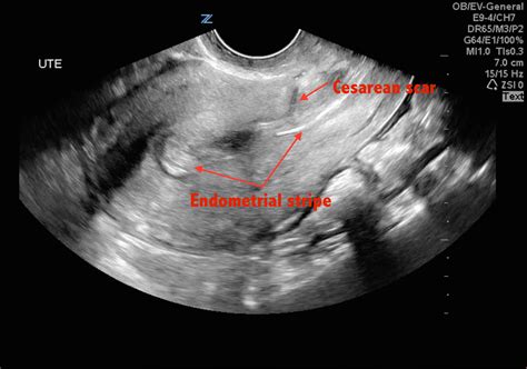
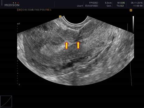
In 2010 Jastrow et al. conducted a meta-analysis of 12 articles on LUS thickness and risk of uterine scar defect and showed a strong association between the . lower uterine segment, Cesarean section, ultrasound and uterine defect (Appendix 1). We searched without language restrictions. . In this Review we describe the value of LUS thickness . Introduction. A wedge-shaped hypoechoic Cesarean section (CS) scar was first described using hysterosalpingography in 1961 1, transabdominal sonography (TAS) in 1982 2 and transvaginal sonography (TVS) in 1990 3.The first two studies were in non-pregnant women and the third included both pregnant and non-pregnant populations.The study showed that ultrasound measurement of 3D ultrasound thick scar on the uterus after previous cesarean section has practical application in determining the mode of delivery among pregnant women who have previously given birth by Caesarean section. . Evaluation of scar thickness is done by ultrasound, but it is still debatable size of .
Based on scar thickness, cases were assigned into two groups of ≤ 3 mm and > 3 mm. TOLAC (Trial of labor after caesarean) was given to cases having scar thickness > 3 mm and elective LSCS was .
All participants underwent an evaluation of uterine scar by using transvaginal ultrasound at 11 to 13 weeks, including the presence of a scar defect and measurement of RMT; and a second evaluation at 35 to 38 weeks, combining both transvaginal and transabdominal ultrasound, for the measurement of LUS thickness. If a niche with a depth of at least 1 mm could be detected, the same measurements as mentioned above were repeated in the distended uterus. In women without a niche, the thickness of the myometrium at the site of the Cesarean scar (if visible) was measured and recorded as the thickness of the residual myometrium.Objectives: To compare residual myometrial thickness (RMT) and size of the Cesarean scar defect after single- and double-layer uterotomy closure following first elective Cesarean section. Methods: A retrospective cohort study was conducted in 149 women at least 6 months after an uncomplicated, elective Cesarean delivery. Two-dimensional transvaginal ultrasonographic .Early diagnosis and appropriate management of Cesarean scar pregnancy (CSP) are crucial to prevent severe complications, such as uterine rupture, severe hemorrhage and placenta accreta spectrum disorders. In this article, we provide a step-by-step tutorial for the standardized sonographic evaluation .
This was a prospective cohort study of women with a singleton pregnancy and a single prior low-transverse CS. All participants underwent an evaluation of uterine scar by using transvaginal ultrasound at 11 to 13 weeks, including the presence of a scar defect and measurement of RMT; and a second evaluation at 35 to 38 weeks, combining both .
Objectives: Lower uterine segment (LUS) thickness measurement using transabdominal ultrasound (TA-US), transvaginal ultrasound (TV-US), or the combination of both methods can detect scar defect in women with prior cesarean. We aimed to compare the sensitivity of three approaches. Methods: Women with prior cesarean underwent LUS thickness measurement . Many authors have tried to utilize transabdominal and transvaginal 2-D ultrasound to measure the scar thickness and detect the healing defects. . The cesarean scar was assessed using the .The risk of bias and concerns regarding applicability were low among most studies. The sonographic measurement was correlated with either delivery outcome or lower uterine segment thickness at the time of repeat cesarean section. The cut-off value for lower uterine segment thickness ranged from 1.5 to 4.05 mm across all studies.
Patient should not be in labour at the time of scar thickness measurement. The degree of fullness of the urinary bladder affects the thickness of the LUS measurement. . Prospective Analysis of Routine Ultrasound Screening of Cesarean Scars, Chandler Mohan, M.D. Carlos Torres, M.D. B. Denise Raynor, M.D. ,Presented at the 40th Annual Clinical .
KEY WORDS: Cesarean section, one layer, scar thickness, two layers Access this article online Quick Response Code site: www.jbcrs.org . The knowledge of this ultrasound measurement
Lower uterine segment (LUS) thickness measurement using transabdominal ultrasound (TA-US), transvaginal ultrasound (TV-US), or the combination of both methods can detect scar defect in women with prior cesarean. We aimed to compare the sensitivity of three approaches. Methods Data are limited with regards to the use of three-dimensional ultrasound imaging in the diagnosis of cesarean scar niche. 14-17 Small studies looking at interobserver reporting of scar niches and its effectiveness have conflicting results. 14-16 A study of 58 women 6 to 15 months following a lower-segment cesarean section found that there was a . Caesarean scar defect (CSD) seriously affects female reproductive health. In this study, we aim to evaluate uterine scar healing by transvaginal ultrasound (TVS) in nonpregnant women with cesarean section (CS) history and to build a predictive model for cesarean scar defects is very necessary. A total of 607 nonpregnant women with previous CS .

The CSD severity is established through measurement of the ratio between myometrial thickness at the scar level and the thickness of adjacent myometrium: . Syrop CH (2003) Detection of cesarean scars by transvaginal ultrasound. Obstet Gynecol. 101:61–65. PubMed Google Scholar Introduction. Cesarean section (CS) is one of the most frequent abdominal surgical operations carried out in the UK 1.The CS rate increased from 12% to 29% in the UK 2 and from 21.2% to 30.1% in the USA 3 between 1990 and 2008. The increasing CS rate and its associated complications has stimulated an interest in the behavior of CS scars and their associated . Purpose The objective of this study was to evaluate whether scar thickness measured by transvaginal sonography and the sequential change in scar thickness from second to third trimester has any association with mode of delivery in patients with previous cesarean. Methods Pregnant women with previous one cesarean section underwent transvaginal .
Evaluation of scar thickness is done by ultrasound, but it is still debatable scar thickness that would be guiding cut-o value for the completion of the delivery by vaginal method (J Dodd et al., 2004) (1). In recent years, the most common indication for cesarean section is the previous cesarean section delivery. New ultrasound grading system for cesarean scar pregnancy and its implications for management strategies: An observational cohort study PLoS One. 2018 Aug 9 . Grade I CSP indicated the GS embedded in less than one-half thickness of the lower anterior corpus; and grade II CSP represented the GS extended to more than one-half thickness of . According to the quality of the healed scar, pregnant women were categorized for the mode of delivery into either a trial for VBAC (if LUS is >2mm and in the absence of other indications for CS) or an elective repeated cesarean section (ERCS) (if LUS thickness is <2mm, the presence of ballooning, funneling, or defects in the LUS, the presence .
ultrasound for cesarean section scar
DOI: 10.29271/jcpsp.2018.05.361 Corpus ID: 13809585; Ultrasound Predictability of Lower Uterine Segment Cesarean Section Scar Thickness. @article{Tazion2018UltrasoundPO, title={Ultrasound Predictability of Lower Uterine Segment Cesarean Section Scar Thickness.}, author={Shazia Tazion and Maimoona Hafeez and Rukhsana Manzoor and Tashhir Rana}, .

vacuum bag drop test
vacuum chamber drop test
WEBRemova Fundos de Imagens: 100% automático, em 5 segundos e com apenas um clique. Existem umas 20 milhões de atividades mais interessantes do que remover fundos de imagens à mão. Graças à inteligência artificial do remove.bg, você pode reduzir o seu tempo de edição - e divertir-se mais! Integração com seu software de workflow.
ultrasound measurement of cesarean scar thickness|ultrasound for cesarean section