macular choroidal thickness measurement|choroidal thickness in adults : traders The choroidal thickness was measured by EDI-OCT subfoveally, and 1500 μm and 3000 μm nasal and temporal to the fovea. Results: Mean age was 34.6 ± 9.8 years . Resultado da Clash of Clans | Portal de suporte da Supercell. Alto volume de mensagens e tempo de espera demorado no suporte. Procuramos responder às perguntas mais comuns na nossa seção de perguntas frequentes, então sempre vale a pena ver se as informações de que precisa já estão lá. A maneira mais .
{plog:ftitle_list}
3 dias atrás · Martin Freeman e Jenna Ortega em cena de Miller's Girl (2024) — Foto: Divulgação . Veja Mais . Agora, os coordenadores de intimidade só poderão falar .
Automated segmentation of SD-OCT data by graph theory and dynamic programming is a fast, accurate, and reliable method to delineate the choroid. This approach will facilitate longitudinal studies evaluating changes in choroid thickness in response to novel optical corrections and in .The choroidal thickness was measured by EDI-OCT subfoveally, and 1500 μm and .Two macular volumetric scans (25×30°) were taken from 30 eyes of 30 young .The average choroidal thickness in the fovea was defined as average thickness .
The choroidal thickness was measured by EDI-OCT subfoveally, and 1500 μm and 3000 μm nasal and temporal to the fovea. Results: Mean age was 34.6 ± 9.8 years . Two macular volumetric scans (25×30°) were taken from 30 eyes of 30 young adult subjects in two sessions, one hour apart. A single rater manually delineated choroid . Choroidal thickness of the right eye was measured by both devices. The central foveal choroidal thickness (CFCT) measured by SD-OCT and SS-OCT was 273.24 ± 54.29 .Automated segmentation of SD-OCT data by graph theory and dynamic programming is a fast, accurate, and reliable method to delineate the choroid. This approach will facilitate longitudinal .
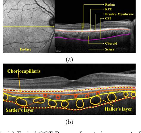
The average choroidal thickness in the fovea was defined as average thickness in the central area of 1000 μm in diameter, according to the Early Treatment Diabetic . Introduction. With the advent of enhanced depth imaging (EDI) technique, 1 manual measurement of subfoveal choroidal thickness based on EDI optical coherence tomography (OCT) images has become a widely used method to investigate choroidal thickness in various macular disorders. In particular, a thin choroid in eyes with exudative and nonexudative age . Purpose: To observe the changes in retinal and choroidal microstructures in patients with different stages of diabetic retinopathy (DR) and to evaluate the vascular perfusion of retina and choroid retinal thickness, retinal and choroidal vessel density by the swept-source optical coherence tomography angiography (SS-OCTA). Methods: Subjects were divided into . Both images were obtained on the same day. The subfoveal choroidal thickness was measured as the distance between the outer border of the retinal pigment epithelium-Bruch’s membrane complex and the chorioscleral border under the fovea. Subfoveal choroidal thickness was 588 μm in EDI-OCT (Top), and 587 μm in SS-OCT (Bottom).
Retinal and Choroidal Vascular Perfusion and Thickness Measurement in Diabetic Retinopathy Patients by the Swept-Source Optical Coherence Tomography Angiography Tingting Liu 1,2,3,4 * † Wei Lin 5 † Genggeng Shi 1,2,3 Wenqi Wang 6 Meng Feng 5 Xiao Xie 6 Tong Liu 7 Qingjun Zhou 2,3,4,8 * Validation of Macular Choroidal Thickness Measurements from Automated SD-OCT Image Segmentation September 2016 Optometry and vision science: official publication of the American Academy of .Purpose.: We compared the reproducibility and mutual agreement of the subfoveal choroidal thickness measurements by expert raters and an automated algorithm in enhanced depth imaging optical coherence tomography (EDI-OCT) images of eyes with nonneovascular age-related macular degeneration (AMD). Methods.: We recruited 44 patients with .
Our data included the basic demographic information of the subjects, the lesion area, perimeter, acircularity index (AI) and VD of FAZ. We also evaluated the changes of retinal and choroidal VD, vascular perfusion, and retinal thickness of the deep vascular complex in the circular radii of 3, 6, 9, and 12 mm around the fovea.
thickness of choroids
Background Macular edema is a common cause of visual loss at uveitic patients. The aim of our study was to investigate retinal and choroidal thickness at the macula in anterior (AU) and intermediate (IMU) uveitis and in healthy individuals using spectral domain optical coherence tomography (SD-OCT). Methods Case-control study of 21 patients with AU and 23 .Spectral domain optical coherence tomography (SD-OCT) is a noninvasive imaging modality that is used commonly to assess retinal thickness, volume, and morphology in pathologic eyes. 1,2 However, evaluating the choroid using standard conventional SD-OCT often is difficult due to the limited signal transmission of the choroidal layer. Recently, the enhanced depth imaging . The choroidal thickness cutoff points that provided the greatest odds of differentiating MMD from non-MMD eyes were an outer nasal sector measuring less than 74 µm (OR=33.8), inner superior sector measuring less than 120 µm (OR=31.4), outer superior sector measuring less than 120 µm (OR=28.2), inner nasal sector measuring less than 85 µm .
To identify the relationship of macular outward scleral height (MOSH) with axial length (AL), macular choroidal thickness (ChT), peripapillary atrophy (PPA), and optic disc tilt in Chinese adults.Whereas the choroid was noted to be significantly thickened in patients with chronic central serous choroioretinopathy 8 –10 and Vogt–Koyanagi–Harada disease, 11,12 it was observed to be significantly thinner in patients with pathologic myopia, 13,14 age-related macular degeneration, 15 –17 age-related choroidal atrophy, 18 glaucoma, 19,20 and diabetic retinopathy. 7,16 . Measurement of choroidal thickness using the proposed in this work for segmentation of the retina and choroid layer. (a) Focal . Berntsen D.A. Validation of macular choroidal thickness measurements from automated SD-OCT image segmentation. Optom. Vis. Sci. 2016; 93:1387. doi: 10.1097/OPX.0000000000000985. [PMC free article] [Google . The average choroidal thickness in age-matched controls was 213.88 ± 45.60 µm. Choroidal thickness increases with the severity of edema; choroidal thickness was higher in diffuse macular edema than in FME. However, choroidal thickness increased in cystic macular edema with subretinal fluid (CME+).
Statistical analysis was used to correlate inter-observer findings, choroidal thickness and area measurements with age, and choroidal thickness with retinal foveal thickness. Results: The 34 subjects had a mean age of 51.1 years. Reliable measurements of choroidal thickness were obtainable in 74% of eyes examined. Purpose The purpose of this study is to assess macular choroidal thickness measurements in patients with different severities of obstructive sleep apnea syndrome (OSAS) versus normal controls by using enhanced depth imaging optical coherence tomography (EDI-OCT). Design This paper is a descriptive study. Materials and methods In this prospective . Our study overcomes the limitations of manual estimations by employing AI to precisely measure the choroidal vascular layer thickness in patients with wet AMD. This novel, AI-based auto-measurement method is accurate, efficient, and detailed. . Choroidal thickness in age-related macular degeneration. Retina, 34 (2014), pp. 1149-1155. View in . Purpose: To compare macular choroidal thickness between patients older than 65 years with early atrophic age-related macular degeneration (AMD) and normals. Methods: This was a consecutive, cross-sectional observational study. Enhanced depth imaging spectral-domain optical coherence tomography using horizontal raster scanning at 12 locations .
To measure the choroidal thickness by enhanced depth imaging optical coherence tomography (EDI-OCT) in normal eyes. In a prospective case series, 208 eyes of 104 normal Iranian subjects were enrolled. . Measurement of retinal thickness is important in the diagnosis, follow-up, and monitoring response to treatment of a number of eye diseases .Variability of subfoveal choroidal thickness measurements in patients with age-related macular degeneration and central serous chorioretinopathy. Eye (Lond). 2013;27(7):809-815. Sigler EJ, Randolph JC. Comparison of macular choroidal thickness among patients older than age 65 with early atrophic age-related macular degeneration and normals. The reliability of retinal and choroidal thickness measurements was very good (all ICC ≥0.98). The variance ratio (F statistic) and the 95% LoA of the intraobserver measurement differences were calculated and were not significantly different for any of the retinal or choroidal thickness parameters . Download: .
choroidal thickness normal eyes
segmentation method to quantify choroid thickness compared to manual segmentation. Methods Two macular volumetric scans (25 × 30°) were taken from 30 eyes of 30 young adult subjects in two sessions, 1 hour apart. A single rater manually delineated choroid thickness as the distance between Bruch’s membrane and sclera across three B-scans (foveal, inferior, and superior . INTRODUCTION. There is a growing interest in the role of the choroid in various chorioretinal diseases. Abnormalities of the choroidal integrity have been associated with the pathogenesis of several retinal diseases, such as age-related macular degeneration (AMD) [1], central serous chorioretinopathy (CSCR) [2] and diabetic maculopathy [3]. New optical . SS-OCT system at 1 μm provides macular choroidal thickness maps and allows one to evaluate the choroidal thickness more accurately. . Three-dimensional volumetric measurement comprised of 512 × 128 A scans was acquired in 0.8 second. From a series of OCT images, a chroidal thickness map of the macular area was created by manual . Methods. A masked, cross-sectional, and consecutive study involving patients with emmetropia/myopia (control group) and high myopia (HM) eyes. Automated choroidal thickness (CT) and manual outer retinal layer (ORL) thickness were acquired using swept-source optical coherence tomography, while retinal sensitivity (RS) assessed by microperimetry (MP3) in all .
Purpose.: To determine choroidal thickness (CT) profile in a healthy population using swept-source optical coherence tomography (SS-OCT). Methods.: This was a cross-sectional, noninterventional study. A total of 276 eyes (spherical equivalent ±3 diopters [D]) were scanned with SS-OCT. Horizontal CT profile of the macula was created measuring subfoveal .
These participants underwent OCT and ophthalmological evaluations, encompassing retinal and choroidal thickness measurements, refractive error, axial length (AL), and systemic examinations. The .

choroidal thickness in women
choroidal thickness in adults
WEB29 de nov. de 2023 · Martina olvr ( COMPLETO DELA E MAIS 200 MODELOS LINK NOS COMENTÁRIOS ) GRUPO NO TELEGRAM AS MELHORES E FAMOSAS MODELOS .
macular choroidal thickness measurement|choroidal thickness in adults