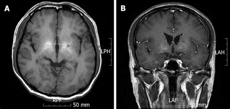
this test uses magnetic waves to show soft tissue lesions|magnetic resonance imaging results : convenience store An MRI scan may be used to look at bones, joints, and soft tissues, such as cartilage, muscles, and tendons for things like: Injuries, such as fractures or tears to a tendon, ligament, or . Resultado da Simply use our menu to navigate yourself through the Sport lemon sports streams and then just click on the fromhot stream link you’ve decided for. That’s it! Watch the Best Live Sports Streams on the internet with Sportlemon! Sportlemon.tv is here to provide you with tons of Live Sports Streams for free!
{plog:ftitle_list}
web26 de ago. de 2023 · Resultado do jogo Mineros de Guayana x Zamora ao vivo (Campeonato Venezuelano) em 26/08/2023. Mineros de Guayana e Zamora com placar .

While both tests show images of structures of your body, an MRI is better at showing contrast and details of soft tissue like the brain, muscles, tendons, ligaments, nerves, and spinal cord, while a CT scan is typically better .Magnetic resonance imaging (MRI) is a diagnostic exam that uses a combination of a large magnet, radiofrequencies and a computer to produce detailed images of organs and structures within the body. MRI does not use ionizing radiation.
magnetic resonance imaging results
An MRI scan may be used to look at bones, joints, and soft tissues, such as cartilage, muscles, and tendons for things like: Injuries, such as fractures or tears to a tendon, ligament, or .
The images reveal abnormalities in both bone and soft tissues, such as pneumonia in the lungs, tumors in different organs, or bone fractures. What does an MRI show? MRI also creates detailed pictures of areas inside . MRI or magnetic resonance imaging uses strong magnetic fields and radio waves to make images of the organs, cartilage, tendons, and other soft tissues of the body. MRI costs .The MRI tool uses magnetic fields and a sophisticated computer to take high-resolution pictures of your bones and soft tissues. In this cross-section MRI scan of a tibia (shinbone), a bone tumor shows up clearly as bright white against .
Magnetic resonance imaging (MRI) of the body uses a powerful magnetic field, radio waves and a computer to produce detailed pictures of the inside of your body. Doctors may use it to help .Magnetic resonance imaging, or MRI, uses a magnet to examine the inside of your body, useful for diagnosing conditions like soft tissue sarcoma. An MRI is a test that uses powerful magnets, radio waves, and a computer to make detailed pictures of the inside of your body. It's helps a doctor diagnose a disease or injury.An X-ray won’t show subtle bone injuries, soft tissue injuries or inflammation. However, even if your doctor suspects a soft tissue injury like a tendon tear, an X-ray might be ordered to rule out a fracture. What injuries require an MRI? .
Magnetic resonance imaging (MRI) is another diagnostic imaging technique that produces cross-sectional images of your body. Unlike X-rays and CT scans, MRI scans work without radiation. The MRI tool uses magnetic fields and a . Magnetic resonance imaging (MRI) is a diagnostic technique useful for noninvasive visualization of organs and soft tissue structures.[1] The ability to evaluate for structural integrity lends MRI to imaging the neural axis and large joints of the musculoskeletal system, where it was used most heavily during its infancy. Since then, MR's scope and .
Soft-tissue lumps and bumps are a common referral for imaging in children and adolescents. The etiology of these lesions includes benign non-tumorous lesions, as well as benign and malignant tumors. Some of these lesions have a characteristic imaging appearance but others do not and require tissue s .Study with Quizlet and memorize flashcards containing terms like lesion, EEG, CT scan and more. . a technique that uses magnetic fields and radio waves to produce computer-generated images that distinguish among different types of soft tissue; allows us to see structures within the brain. brainstem.Using a magnetic field and radio waves, an MRI scan creates detailed three-dimensional images of the structures in your body. . This test allows your doctor to determine whether a soft tissue sarcoma has developed from muscle, fat, or other tissue. . our doctors may look for changes in the chromosomes—the portions of cells that house .A magnetic resonance (REZ-oh-nans) imaging scan is usually called an MRI. An MRI does not use radiation (X-rays) and is a noninvasive medical test or examination. The MRI machine uses a large magnet and a computer to take pictures of the inside of your body. Each picture or "slice" shows only a few layers of body tissue at a time.
Magnetic Resonance Imaging (MRI) is a non-invasive imaging technology that produces three dimensional detailed anatomical images. It is often used for disease detection, diagnosis, and treatment monitoring. It is based on sophisticated technology that excites and detects the change in the direction of the rotational axis of protons found in the water that makes up living tissues. Musculoskeletal soft tissue tumors are a heterogeneous group including tumor-like lesions, benign, and malignant tumors [] with a benign to malignant ratio of over 100:1 [].An overall incidence of 300 cases per 100,000 population has been reported [].Whereas it is essential to accurately identify the potentially malignant lesions for timely therapy, the imaging .
What are benign soft tissue tumors? Benign soft tissue tumors are noncancerous lumps under your skin.They develop anywhere you have soft tissue such as your muscles, tendons and fat. Depending on your situation, your healthcare provider may recommend surgery to remove the tumor and/or radiation therapy to keep the tumor from coming back (recurring).
MRI Imaging for Osseous Lesions. MRI uses strong magnetic fields and radio waves to generate detailed images of the bones and surrounding soft tissues. MRI is particularly useful for identifying the involvement of surrounding soft tissues, marrow, and cartilage. It helps in determining the exact nature and extent of the lesions. When to See a .Magnetic resonance imaging (MRI) of the body uses a powerful magnetic field, radio waves and a computer to produce detailed pictures of the inside of your body. . MR images of the soft-tissue structures of the body—such as the liver and many other organs—are sometimes more likely to accurately identify disease than other imaging methods . Objective To predict accurately whether a soft tissue mass was benign or malignant and to characterize its type using ultrasound, shear wave elastography and MRI. We hypothesized that with the addition of shear wave elastography, it would be possible to determine a threshold velocity value to classify a lesion as benign or malignant. Materials and methods .
a technique that uses magnetic fields and radio waves to produce computer-generated images that distinguish among different types of soft tissue; allows us to see structures within the brain. positron emission tomography (PET) scanThis magnetic field, along with a radio wave, briefly redirects the hydrogen atoms' natural alignment in the body. . Cross-sectional views can be done to show more details. MRI does not use ionizing radiation, like X-rays or CT scans. . , joints, or soft tissue. What are the risks of an MRI scan? There is no risk of exposure to radiation .
magnetic resonance imaging procedures
MRI, or Magnetic Resonance Imaging, is a non-invasive medical imaging technique that uses powerful magnetic fields and radio waves to generate detailed images of the body's internal structures. In the case of brain .Study with Quizlet and memorize flashcards containing terms like Tissue distruction, An amplified recording of the waves of electrical activity sweeping across the brain's surface. These waves are measured by electrodes placed on the scalp., A brain-imaging technique that measures magnetic fields from the brain's natural electrical activity. and more.
An MRI provides superior tissue contrast resolution. Because of its ability to show soft tissues in exquisite detail, MRI can detect soft tissue disease and evaluate vasculature. An MRA, or magnetic resonance angiogram, is a special type of MR that creates three-dimensional reconstructions of vessels containing flowing blood. An ultrasound is one of several tests that healthcare professionals might use to confirm a diagnosis of soft tissue sarcoma. This type of imaging uses high frequency sound waves to create detailed .
Introduction. Soft tissue malignancies are an uncommon heterogeneous group of mesenchymal lesions. They account for 1% of adult malignant tumors 1–3 and are estimated to represent about 1% of all malignant tumors with a lifetime risk of development estimated at 0.33%. 4. Long-term local and systemic disease-free survival depends on patient age and .Tissue destruction. Brain lesions occur naturally (disease or trauma), in surgery, or experimentally (using electrodes to destroy brain cells). . A technique that uses magnetic fields and radio waves to produce computer-generated images of soft tissue. MRI scans show brain anatomy. fMRI (functional MRI)
The most recent study suggested that malignant soft tissue lesions show variable elasticity according to cellular differentiation and tissue characteristics. Therefore, more studies should be done to identify ways of differentiating malignant soft tissue tumors from benign tumors [11-14]. However, most soft tissue tumors are benign lesions. 1. Introduction. Soft tissue tumors (STT) are represented by a wide range of different histological and molecular subsets with very low incidence populations at all ages [].STT represents the majority of sarcomas (soft tissue tumors ~75%, gastrointestinal ~15%, and ~10% bone sarcomas) [].The prognosis of STT is influenced by different factors such as grading, . An MRI is a test that uses powerful magnets, radio waves, and a computer to make detailed pictures of the inside of your body. It's helps a doctor diagnose a disease or injury.MRI uses magnetic fields and radio waves to produce images of thin slices of tissues (tomographic images). Normally, protons within tissues spin to produce tiny magnetic fields that are randomly aligned. . T1-weighted images optimally show normal soft-tissue anatomy and fat (eg, to confirm a fat-containing mass). T2-weighted images optimally .
magnetic resonance imaging problems
medidor de umidade de grãos moisture-max plus
Soft Tissue Masses: Ultrasound is a useful tool for the identification and characterization of soft tissue masses, including but not limited to lipomas (soft, benign tumors made of fatty tissue), cysts (sac-like structures filled with fluid or semi-solid material), and abscesses (pockets of pus caused by infection or inflammation).
Magnetic resonance imaging (MRI) uses powerful magnetic forces and radiofrequency waves to make cross-sectional images of organs, tissues, bones and blood vessels. A computer turns the images into 3D pictures. An MRI is commonly used to examine a lump that doctors think may be a soft tissue sarcoma.
medidor de umidade de grãos moisture-max plus preco
medidor de umidade de grãos motomco 999fr preço
Resultado da Wolves have been known to disperse up to 550 miles, but more commonly disperse 50 – 100 miles from their natal pack. Generally wolves disperse when 1 – 2 years old as they reach sexual maturity although some adults disperse also. At any one time 5 – 20 percent of the wolf population may be .
this test uses magnetic waves to show soft tissue lesions|magnetic resonance imaging results